
Z-VAD-FMK: Mechanism of Action, Working Concentration, and How It Compares to Other Caspase Inhibitors
How the pan-caspase inhibitor Z-VAD-FMK works, what working concentration to use, how to design controls, and how it compares with Q-VD-OPh, Boc-D-FMK and selective caspase inhibitors.
Technical guide from Adooq Bioscience · Product: Z-VAD-FMK, Cat. No. A12373 · Last reviewed: November 2026
Z-VAD-FMK at a glance
| Property | Value |
|---|---|
| Common name | Z-VAD-FMK (sold as the methyl ester, Z-VAD(OMe)-FMK) |
| Full name | benzyloxycarbonyl-Val-Ala-Asp(OMe)-fluoromethylketone |
| CAS | 187389-52-2 |
| Molecular formula | C22H30FN3O7 |
| Z-VAD-FMK molecular weight | 467.49 g/mol |
| Free-acid form | Z-VAD(OH)-FMK, C21H28FN3O7, MW 453.46, CAS 161401-82-7 |
| Mechanism | Cell-permeable, irreversible pan-caspase inhibitor. The fluoromethyl ketone alkylates the catalytic cysteine |
| Caspases covered | Caspase-1, -3, -4, -5, -6, -7, -8, -9, -10, -11 (caspase-2 is inhibited poorly) |
| Solubility | DMSO 80 mg/mL (171 mM); insoluble in water and ethanol |
| Typical stock | 10-50 mM in anhydrous DMSO, single-use aliquots, -20 °C |
| Z-VAD-FMK working concentration | 20-50 µM for most cell culture; 10-100 µM is the published range |
| Pre-treatment | 30-60 min before the death stimulus |
| In vivo vehicle | 2% DMSO / 35% PEG 300 / 2% Tween 80 / ddH2O, up to about 5-6 mg/mL |
| Storage | -20 °C desiccated, protected from light; DMSO stocks for up to a month at -20 °C |
Z-VAD-FMK is the broadest and cheapest way to ask whether a phenotype is caspase-dependent. It is also the fastest way to convert apoptosis into necroptosis by accident, which is why most of this guide is about interpretation rather than pipetting.
1. What Z-VAD-FMK is
Z-VAD-FMK is a tripeptide caspase inhibitor built from three functional parts, and every practical property of the molecule falls out of one of them.
The "Z" cap (benzyloxycarbonyl). It blocks the N-terminus, removes the free amine, and makes the peptide neutral and lipophilic enough to cross the plasma membrane. Unprotected peptides do not get into cells.
The "VAD" recognition element (Val-Ala-Asp). Caspases are the only mammalian proteases that absolutely require aspartate at the P1 position, which is what makes a tripeptide ending in Asp a caspase-directed reagent at all. VAD is a deliberately generic P4-P2 sequence. It is not the optimal sequence for any single caspase, which is exactly why it binds most of them. The combinatorial substrate-preference mapping of the caspase family by Thornberry and colleagues showed that P4-P2 preferences differ sharply between the three caspase subgroups: DEVD for the executioners, (I/L)EXD for the initiators, WEHD/YVAD for caspase-1 (Thornberry et al. 1997). A tripeptide that avoids all three optima ends up as a broad-spectrum binder.
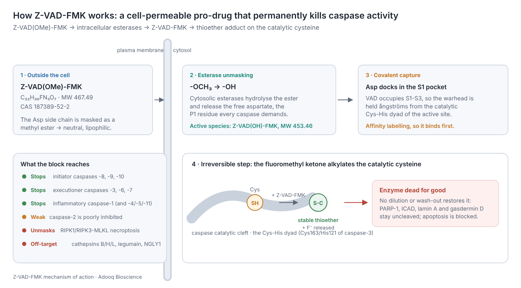
The "FMK" warhead (fluoromethyl ketone). The warhead does the actual inactivating. The ketone carbon is attacked by the catalytic cysteine thiolate, the fluoride is displaced, and the result is a stable thioether, which is a permanent covalent modification of the enzyme. Peptide methyl ketones were characterised as mechanism-based inactivators of interleukin-1β converting enzyme (caspase-1) before the caspase family was even named, when Thornberry and colleagues worked out the kinetics for the (acyloxy)methyl ketone series (Thornberry et al. 1994). Halomethyl ketones such as FMK inactivate by the same alkylation chemistry.
Z-VAD-FMK vs Z-VAD(OMe)-FMK: the same compound, almost
This is the single most common point of confusion, and the reason the keyword "z vad ome fmk" exists at all.
The aspartate side chain carries a carboxylate. A free carboxylate is charged at physiological pH and crosses membranes badly. So essentially every commercial "Z-VAD-FMK", including Adooq A12373, is supplied as the methyl ester, Z-VAD(OMe)-FMK (MW 467.49). Once inside the cell, non-specific cytosolic esterases hydrolyse the ester and liberate the free acid, which is the species that engages the caspase active site.
Practical consequences:
- Use the OMe form for cells and animals. It is the standard, and it is what the published 20-50 µM protocols were built on.
- Use the free acid, Z-VAD(OH)-FMK (MW 453.46), for cell-free work. In purified caspase assays, lysate experiments and isolated mitochondria there are no esterases to do the unmasking, and the ester would simply sit there.
- Watch your molar maths. 467.49 versus 453.46 is only a 3% difference, but people routinely weigh out one form and calculate with the other's molecular weight.
- Esterase activity varies between cell types and is lower in serum-free medium, which is one reason the effective concentration is not identical in every line.
2. Z-VAD-FMK mechanism of action
2.1 How the covalent block works
Reversible inhibitors sit in the active site and come back off, so their effect is governed by Ki and by how long the drug stays above that Ki. Z-VAD-FMK works differently. It is an affinity label: the peptide part provides the address, and once bound, the warhead forms a covalent bond with the catalytic cysteine of the Cys-His dyad. That residue is Cys163 of caspase-3 in UniProt numbering, and you will also see it called Cys285 in the structure-based numbering carried over from caspase-1.
Three bench-level consequences follow:
- Potency depends on exposure time as well as concentration. The right parameter for an irreversible inhibitor is the second-order inactivation rate (kinact/Ki), which is how Garcia-Calvo and colleagues reported their systematic comparison of peptide-based caspase inhibitors (Garcia-Calvo et al. 1998). An IC50 quoted for Z-VAD-FMK is only meaningful next to the pre-incubation time used to measure it, which is why published IC50 values for this compound vary across orders of magnitude and why vendor IC50 tables for it should be read with suspicion.
- Washing it out does not restore caspase activity. Modified enzyme stays modified, and recovery requires new protein synthesis. That helps in long experiments and hurts if you wanted a reversible, titratable block.
- The block is stoichiometric and consumable. Each inhibitor molecule kills one caspase molecule and is spent. In cells that keep making pro-caspases, or that activate a large pool rapidly as in an inflammasome response, the inhibitor can be exhausted. This is why a single dose at t = 0 often fails in 48-72 h experiments and why re-dosing every 12-24 h is standard practice.
2.2 What "pan-caspase" really means
Z-VAD-FMK inhibits initiator caspases (-8, -9, -10), executioner caspases (-3, -6, -7) and inflammatory caspases (-1, -4, -5, and murine -11). Coverage is uneven. Caspase-2 is notoriously poorly inhibited, and relative potency against the other family members differs by more than an order of magnitude. "Pan" means the compound hits most caspases, at different rates.
The corollary for experimental design: failure to rescue with Z-VAD-FMK is not proof that caspases are uninvolved. It may mean caspase-2, insufficient concentration, insufficient pre-incubation, or an exhausted inhibitor pool.
2.3 What is downstream of the block
When caspases are inactivated, the hallmark events of apoptosis do not occur:
- PARP-1 is not cleaved to its 89 kDa fragment
- ICAD/DFF45 is not cleaved, so CAD stays inhibited and the DNA ladder and TUNEL signal do not appear
- Lamin A and the cytoskeletal substrates survive, so nuclear fragmentation and blebbing are suppressed
- Gasdermin D is not processed by caspase-1, so pyroptotic pore formation and mature IL-1β release are suppressed
- Phosphatidylserine exposure (annexin V) is delayed or lost, because flippase and scramblase control is caspase-dependent
These double as your positive controls that the inhibitor is working. If PARP cleavage is unchanged on a western blot, check the stock, the pre-incubation and the concentration before you conclude anything biological.
3. Why researchers use Z-VAD-FMK: five distinct jobs
It helps to be explicit about which of these five experiments you are running, because the right concentration, timing and controls differ for each.
Establishing caspase dependence. The default use. A stimulus kills cells, Z-VAD-FMK prevents it, therefore the death is caspase-dependent. This is the backbone of thousands of papers.
Rescuing viability to study something else. If you need cells alive long enough to measure a downstream event such as differentiation, viral replication or cytokine release, a pan-caspase block buys you time.
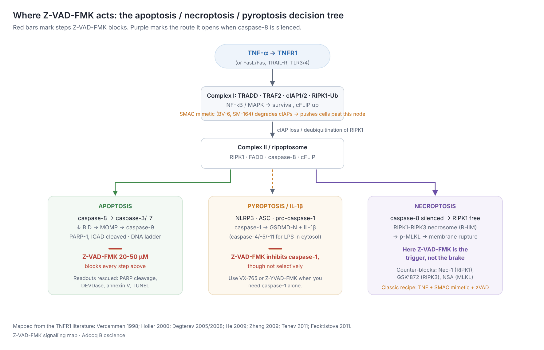
Deliberately unmasking non-apoptotic death. This inverted use is now as important as the first. Blocking caspase-8 removes the brake on RIPK1 and RIPK3, so adding Z-VAD-FMK to a TNF stimulus induces necroptosis instead of preventing death. Vercammen and colleagues showed this in L929 cells in 1998, before the pathway had a name: caspase inhibition increased sensitivity to TNF-mediated necrosis rather than protecting against it (PMID 9565639).
Suppressing inflammasome output. Caspase-1 inhibition blocks gasdermin D cleavage, pyroptosis and mature IL-1β and IL-18 release. Z-VAD-FMK works here, but it is a blunt instrument (see §7).
Preventing secondary necrosis artefacts. In long time-lapse imaging, or in cultures harvested at late time points, apoptotic corpses lyse and release contents that confound LDH, cytokine and nucleic-acid readouts. A caspase block keeps the preparation clean.
4. Experiment types and research areas
4.1 Apoptosis research
The core application, and the reason for the keyword "z vad fmk apoptosis". Standard pairings:
| Death stimulus | What it does | Typical companion |
|---|---|---|
| Staurosporine | Broad kinase inhibition, strong intrinsic apoptosis; the field's positive control | Staurosporine, 0.1-1 µM, 3-6 h |
| TNF-α + cycloheximide | Blocks NF-κB-driven survival protein synthesis, converts TNF into a death signal | CHX 1-10 µg/mL |
| FasL / anti-Fas (CH-11) | Extrinsic, caspase-8-driven | ± CHX |
| TRAIL | Extrinsic, DR4/DR5 | Often with SMAC mimetic |
| Etoposide, cisplatin, doxorubicin | Genotoxic, intrinsic, p53-dependent | 12-48 h exposures |
| UV / γ-irradiation | Intrinsic | n/a |
| Growth factor / serum withdrawal | Intrinsic | n/a |
Typical readouts rescued: caspase-3/7 DEVDase activity, PARP cleavage by western blot, annexin V/PI flow cytometry, TUNEL, nuclear morphology by Hoechst, and MTT or CellTiter-Glo viability.
4.2 Necroptosis research
The canonical induction cocktail is TNF-α plus a SMAC mimetic plus Z-VAD-FMK, often abbreviated TSZ:
- TNF-α engages TNFR1.
- A SMAC mimetic, BV-6 or SM-164, degrades cIAP1/2, destabilising complex I and allowing the cytosolic death complex to assemble.
- Z-VAD-FMK removes caspase-8, which would otherwise cleave and inactivate RIPK1 and RIPK3.
What remains is the RIPK1-RIPK3 necrosome, MLKL phosphorylation, and lytic death. The genetic and pharmacological logic of this pathway was built on exactly this combination: RIP kinase as a caspase-independent death effector (Holler et al. 2000), necrostatin-1 as a RIPK1 inhibitor (Degterev et al. 2005, 2008), and RIPK3 as the switch that decides between apoptosis and necrosis (He et al. 2009; Zhang et al. 2009). The ripoptosome work (Tenev et al. 2011; Feoktistova et al. 2011) explains why IAP antagonism is the third required ingredient, and the RIPK3 kinase-activity work behind GSK'872 (Mandal et al. 2014) supplies the counter-inhibitor. Linkermann and Green's review is the best short orientation to the whole pathway (Linkermann & Green 2014).
Controls to run in necroptosis work:
- Necrostatin-1 (RIPK1) should block the death that Z-VAD-FMK unmasked
- GSK-872 (RIPK3) does the same, one step down
- Necrosulfonamide (MLKL, human cells only) or MLKL knockdown
- p-MLKL and p-RIPK3 western blots alongside the viability data
4.3 Pyroptosis and inflammation
In LPS-primed macrophages or THP-1 cells stimulated with nigericin, ATP or monosodium urate crystals, Z-VAD-FMK blocks caspase-1-dependent IL-1β maturation and pyroptotic lysis. The underlying event, caspase-1-dependent pore formation leading to osmotic lysis of the infected macrophage, was characterised by Fink and Cookson (2006), and a pan-caspase inhibitor will suppress it. The compound is a reasonable first-pass reagent and a poor final answer: because it also blocks caspase-8, which can process pro-IL-1β independently, and caspase-3 and -7, a positive result tells you that a caspase is involved and nothing more. For caspase-1-specific claims, use VX-765/belnacasan or Z-YVAD-FMK alongside it.
4.4 Neuroscience and ischaemia-reperfusion
Caspase activation contributes to delayed neuronal death after ischaemia and excitotoxic injury, and peptide fluoromethyl ketone inhibitors were among the earliest tools used to test that in vivo, typically by intracerebroventricular infusion, because these compounds cross the blood-brain barrier poorly. Hara and colleagues used exactly this approach to show that inhibiting the ICE/caspase family reduces ischaemic and excitotoxic neuronal damage in rodents (Hara et al. 1997), and Van Noorden's review traces the wider history of the compound in vivo (Van Noorden 2001). The BBB limitation is the single best reason to consider Q-VD-OPh, which is described as brain-permeable, for CNS studies (Caserta et al. 2003).
4.5 Virology and infection biology
Two standard uses. First, preventing host-cell apoptosis to let a viral replication cycle complete, which separates "the virus kills the cell" from "the virus needs the cell". Second, in bacterial infection models, distinguishing caspase-1-driven pyroptosis of infected macrophages from apoptotic or necroptotic death. Caspase inhibition during infection frequently redirects the cell toward necroptosis rather than rescuing it. TLR3 engagement, for example, drives RIP3- and MLKL-dependent necrosis through TRIF when caspase-8 is unavailable (Kaiser et al. 2013), so always check.
4.6 Immunology and sepsis
Lymphocyte apoptosis is a major feature of sepsis, and caspase inhibition has been tested in animal models as a way to preserve the lymphocyte compartment. Hotchkiss and colleagues showed that caspase inhibitors improved survival in a mouse sepsis model and that the benefit depended on the lymphocyte compartment (Hotchkiss et al. 2000). Whether blocking apoptosis helps or merely shifts the mode of death is still an open therapeutic question, and it is also why Z-VAD-FMK itself never became a drug while orally bioavailable successors such as emricasan reached human trials.
4.7 Developmental biology, toxicology, cardiology
Programmed cell death during development, drug-induced hepatotoxicity, cardiomyocyte death after ischaemia-reperfusion, and chemotherapeutic mechanism-of-action studies all use the same basic design: does the pan-caspase block rescue the phenotype?
4.8 Cell-death-pathway triage
Z-VAD-FMK is one arm of a four-inhibitor panel used to classify an unknown death.
| Add | If death is blocked, suspect |
|---|---|
| Z-VAD-FMK (20-50 µM) | Apoptosis or pyroptosis |
| Necrostatin-1 (10-30 µM) | Necroptosis (RIPK1-dependent) |
| Ferrostatin-1 / liproxstatin-1 | Ferroptosis |
| 3-MA / bafilomycin A1 | Autophagy-dependent death |
Death that is blocked by Z-VAD-FMK plus Nec-1 only in combination is the classic signature of a system that flips between pathways.
5. Z-VAD-FMK working concentration: a practical protocol
5.1 Stock solution
- Spin the vial before opening. The compound ships as a film or lyophilised powder.
- Dissolve in anhydrous DMSO. Solubility is 80 mg/mL (171 mM), so a 20 mM or 50 mM stock is comfortable. For 1 mg of MW 467.49 material, 107 µL of DMSO gives 20 mM.
- Aliquot single-use volumes immediately and store at -20 °C, or -80 °C for long term. Avoid freeze-thaw cycles: the fluoromethyl ketone is the reactive part of the molecule and it does not improve with handling.
- Use DMSO stocks within about a month. Make the working dilution into medium immediately before use, and never dilute into aqueous buffer for storage.
- Keep final DMSO at or below 0.1% (v/v) and run a vehicle-matched control. At 50 µM from a 20 mM stock, DMSO is 0.25%, so either raise the stock concentration or accept that figure and match it.
5.2 Choosing the concentration
| Application | Z-VAD-FMK concentration | Timing | Notes |
|---|---|---|---|
| Standard apoptosis block (most cell lines) | 20-50 µM | 30-60 min pre-treatment, then co-incubate | The default starting point |
| Sensitive / primary cells, short assays | 10-20 µM | 30 min pre-treatment | Often sufficient; less vehicle load |
| Strong stimuli (staurosporine, high TRAIL), resistant lines | 50-100 µM | 1 h pre-treatment, re-dose at 24 h | Above 100 µM, off-target effects dominate |
| Necroptosis induction (TSZ) | 20-50 µM | Added with or just before TNF-α | Here the block is the stimulus |
| Inflammasome / IL-1β suppression | 20-50 µM | With or shortly after the priming step | Not caspase-1-selective |
| Long experiments (48-72 h) | 20-50 µM, re-dosed every 24 h | n/a | The inhibitor is consumed and degraded |
| Cell-free / lysate caspase assays | 1-100 µM of Z-VAD(OH)-FMK | 15-30 min pre-incubation with enzyme | Use the free acid, not the ester |
| In vivo (rodent, IP) | Formulate at about 5 mg/mL in 2% DMSO / 35% PEG 300 / 2% Tween 80 / water | Dose per the specific model | Doses in the literature vary widely by model and route; see §5.5 |
InvivoGen's technical sheet for the same compound recommends a 1-10 µg/mL working range, which is approximately 2-21 µM, consistent with the lower end of the published range.
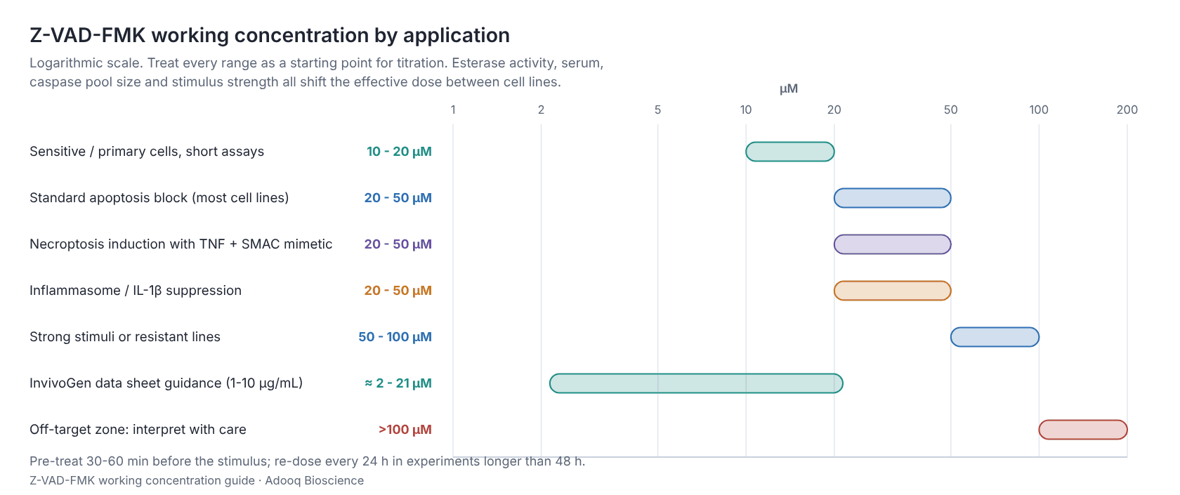
Always titrate in your own system. Reported effective concentrations for the same readout differ several-fold between cell lines, chiefly because of differences in esterase activity, caspase pool size, serum protein binding and stimulus strength.
5.3 Timing rules that matter
- Pre-incubate. Covalent inactivation takes time, and the inhibitor must win the race against caspase activation. 30-60 min before the stimulus is standard, and 2 h for slow-uptake primary cells.
- Do not wash out and expect reversal. To confirm the block is on-target, add inhibitor after caspase activation has begun and show that the block is weaker, rather than looking for reversal on washout.
- Re-dose long experiments. A 72 h experiment dosed only at t = 0 is a common source of "Z-VAD-FMK stopped working".
5.4 Controls you should run
| Control | Purpose |
|---|---|
| DMSO vehicle, concentration-matched | Rules out solvent effects |
| Z-FA-FMK | The standard negative control: same Z-cap and FMK warhead, but no P1 aspartate, so it does not inhibit caspases. It does inhibit cathepsins, which makes it a direct control for the cathepsin off-target problem (see §7) |
| Z-VAD-FMK alone, no stimulus | Detects inhibitor toxicity and spontaneous necroptosis induction |
| PARP / caspase-3 cleavage western | Confirms the block is biochemically real |
| Necrostatin-1 co-treatment | Reveals a pathway switch rather than a rescue |
| Q-VD-OPh in parallel | A different scaffold reaching the same conclusion is far stronger evidence |
5.5 In vivo use
Z-VAD-FMK is active in animals, and three studies define the shape of that literature.
Rodriguez and colleagues showed that systemic injection of a Z-VAD-type tripeptide inhibitor blocked intracellular activation of CPP32-like proteases in vivo and fully protected mice against Fas-mediated fulminant liver destruction (Rodriguez et al. 1996). That remains the cleanest demonstration that the compound reaches its target in a whole animal. In sepsis, caspase inhibition improved survival in a mouse model, with the benefit traced to preservation of lymphocytes (Hotchkiss et al. 2000). In CNS injury, inhibition of the ICE/caspase family reduced ischaemic and excitotoxic neuronal damage, delivered intracerebroventricularly because of poor blood-brain-barrier penetration (Hara et al. 1997).
Practical points:
- Take the dose, route and schedule from the paper closest to your model. Published regimens differ by an order of magnitude between models, species and routes, and a dose transplanted from an unrelated model is a guess rather than a starting point.
- Formulate fresh. A standard IP vehicle is 2% DMSO / 35% PEG 300 / 2% Tween 80 in water, to roughly 5-6 mg/mL. Oral gavage suspensions use 1% CMC-Na.
- Expect poor CNS exposure. For brain work, either dose intracerebroventricularly or switch to Q-VD-OPh.
- Watch the FMK liability. Repeat dosing of a fluoromethyl ketone is the setting where fluoroacetate formation is most often raised as a concern (§7.5), which is another reason groups move to Q-VD-OPh or emricasan for chronic studies.
6. Molecules commonly used alongside Z-VAD-FMK
| Molecule | Role | Why it appears with Z-VAD-FMK |
|---|---|---|
| TNF-α | Death-receptor ligand | The trigger in both apoptosis (with CHX) and necroptosis (with SMAC mimetic + zVAD) |
| Cycloheximide | Protein synthesis inhibitor | Removes short-lived cFLIP and NF-κB survival proteins so TNF can kill |
| BV-6, SM-164, birinapant | SMAC mimetics / IAP antagonists | Degrade cIAP1/2 and permit ripoptosome and necrosome assembly |
| Necrostatin-1 | RIPK1 inhibitor | The counter-block that identifies necroptosis |
| GSK-872 | RIPK3 inhibitor | Second-line necroptosis confirmation |
| Necrosulfonamide | MLKL inhibitor (human) | Terminal-step confirmation |
| Staurosporine | Broad kinase inhibitor | The standard apoptosis positive control |
| TRAIL, FasL, anti-Fas | Death-receptor ligands | Extrinsic-pathway stimuli |
| Etoposide, cisplatin, doxorubicin | Genotoxic agents | Intrinsic-pathway stimuli |
| LPS + nigericin / ATP / MSU | Inflammasome priming and activation | Pyroptosis and IL-1β experiments |
| Z-FA-FMK | Non-caspase FMK peptide | Negative control for caspase specificity, positive control for cathepsin inhibition |
| Ferrostatin-1, 3-MA, bafilomycin A1 | Ferroptosis and autophagy blockers | Pathway triage panel |
| Biotin-VAD-FMK | Activity-based probe | Labels the active caspases that Z-VAD-FMK would have hit |
7. Limitations and off-target effects
7.1 It inhibits cysteine proteases that are not caspases
The fluoromethyl ketone warhead is reactive toward cysteine protease active sites in general. Z-VAD-FMK has been shown to inhibit cathepsins, notably cathepsin B, along with legumain and other papain-family enzymes, in work arguing that for these reagents the reaction mechanism matters more than the peptide's nominal specificity (Schotte et al. 1999; Rozman-Pungercar et al. 2003). At 50-100 µM, lysosomal cysteine protease inhibition is not a hypothetical.
Mitigation: run Z-FA-FMK in parallel. It inhibits cathepsins but not caspases, so a phenotype reproduced by Z-FA-FMK is a cathepsin phenotype.
7.2 It inhibits NGLY1 (peptide:N-glycanase)
Z-VAD-FMK was identified as an inhibitor of cytosolic peptide:N-glycanase (NGLY1), an enzyme of the ERAD and glycoprotein degradation pathway with no connection to cell death (Misaghi et al. 2004). Any experiment involving ER stress, glycoprotein quality control or ERAD should treat this as a live confounder.
7.3 It induces autophagy and, in some lines, kills cells by itself
In L929 fibrosarcoma cells, zVAD-fmk alone induces death with autophagic features, and the relationship between caspase-8, the ATG7 and beclin-1 machinery and this death has been dissected in detail (Yu et al. 2004; Wu et al. 2008, 2011). In practice this means a "Z-VAD-FMK only" arm is not an inert control in every cell line, so always run it.
7.4 It converts apoptosis into necroptosis
This is the most consequential limitation and the most common misinterpretation in the literature. If a death stimulus kills a cell that expresses RIPK3 and MLKL, blocking caspase-8 with Z-VAD-FMK can increase death rather than prevent it. A failed rescue is therefore ambiguous: caspases may be irrelevant, or they may have been so relevant that removing them opened a second route. Resolve it with Nec-1 or GSK-872 co-treatment, rather than arguing from the viability curve alone.
7.5 Fluoroacetate metabolism
Fluoromethyl ketones release fluoride on reaction and can, in principle, generate fluoroacetate, a metabolic poison that blocks aconitase in the TCA cycle. This is a long-standing concern for the FMK class in vivo and over long in vitro incubations (Van Noorden 2001), and avoiding it is one of the explicit design arguments for the O-phenoxy (OPh) warhead in Q-VD-OPh (Caserta et al. 2003).
7.6 Caspase-2 escapes
If your hypothesis involves caspase-2, as in certain genotoxic and metabolic stress pathways, Z-VAD-FMK is the wrong reagent.
7.7 Quoted IC50 values are not comparable
Because inactivation is time-dependent, an IC50 for an irreversible inhibitor is a function of the assay's pre-incubation time. Treat any single IC50 number for Z-VAD-FMK, including vendor numbers, as a rough guide rather than a constant. Chauvier and colleagues made the broader version of this argument directly, asking how much of the "broad-spectrum caspase inhibitor" label survives contact with the data (Chauvier et al. 2007).
8. Similar products: how Z-VAD-FMK compares
8.1 Comparison table
| Compound | Type / selectivity | Reversible? | Advantages vs Z-VAD-FMK | Disadvantages |
|---|---|---|---|---|
| Z-VAD-FMK (A12373) | Pan-caspase tripeptide FMK | Irreversible | Broadest coverage, very large literature base, inexpensive, the field's default comparator | Off-target cathepsin, legumain and NGLY1 inhibition; induces autophagy and necroptosis; weak on caspase-2; potential fluoroacetate; needs 20-50 µM |
| Q-VD-OPh | Pan-caspase, quinoline-Val-Asp-OPh | Irreversible | Effective at roughly 10x lower concentration (caspase-3 IC50 25 nM, caspase-1 50 nM, caspase-8 100 nM, caspase-9 430 nM); much lower cytotoxicity; no fluoromethyl ketone, so no fluoroacetate concern; brain-permeable; better for long incubations and in vivo | More expensive; smaller historical literature; still not caspase-2-complete; still unmasks necroptosis |
| Boc-D-FMK | Pan-caspase, single Asp + FMK | Irreversible | Minimal peptide, cheapest, sequence-agnostic | Lower potency; needs higher concentrations; same FMK off-target liabilities, arguably worse because there is almost no recognition element |
| Z-VAD(OH)-FMK | Free-acid Z-VAD-FMK | Irreversible | The correct choice for cell-free and lysate assays | Poor cell permeability, so not for intact cells |
| Emricasan (IDN-6556) | Pan-caspase, non-peptide-like | Irreversible | Orally bioavailable and taken into randomised human trials in cirrhosis and portal hypertension (Garcia-Tsao et al. 2019, 2020); the right tool when translational relevance matters; combines with SMAC mimetics to drive necroptosis in AML models (Brumatti et al. 2016) | Not a classic bench tool; different PK; more expensive |
| Z-DEVD-FMK | Caspase-3/-7-preferring | Irreversible | Asks a narrower question: is the executioner arm required? | Selectivity is only relative. Caspase cleavage-motif preferences overlap heavily (McStay et al. 2008), so DEVD-directed reagents also hit other caspases at working concentrations |
| Z-IETD-FMK | Caspase-8-preferring | Irreversible | Dissects the extrinsic and death-receptor arm; useful for separating apoptosis from necroptosis initiation | Cross-reacts with caspase-10 and granzyme B-like activities; same caveat on relative selectivity |
| Z-LEHD-FMK | Caspase-9-preferring | Irreversible | Dissects the intrinsic and mitochondrial arm | Cross-reacts with caspase-4 and -5; weakest selectivity of the set |
| Z-YVAD-FMK | Caspase-1-preferring | Irreversible | The FMK option for inflammasome work | Still hits other caspases at the concentrations commonly used |
| Ac-DEVD-CHO | Caspase-3/-7 aldehyde | Reversible | Tight-binding and reversible, so Ki is meaningful; the right tool for enzymology on purified protein or lysates | Poor cell permeability; aldehydes react with other cysteine and serine proteases; not for cell-based rescue |
| VX-765 / belnacasan | Caspase-1 (and -4/-5) selective prodrug | Irreversible | Genuinely selective for the inflammatory arm; orally active, with documented suppression of IL-1β and IL-18 release (Wannamaker et al. 2007) | Narrow by design, so no use for apoptosis questions |
| Biotin-VAD-FMK | Tagged pan-caspase probe | Irreversible | Labels active caspases for pull-down or imaging | A detection reagent rather than an inhibitor for rescue experiments |
| Inhibitor | Casp-1 | Casp-2 | Casp-3/-7 | Casp-6 | Casp-8/-10 | Casp-9 | Cathepsins | NGLY1 | Cell permeant |
|---|---|---|---|---|---|---|---|---|---|
| Z-VAD-FMK (OMe) | Strong | Weak | Strong | Strong | Strong | Strong | Off-target | Off-target | Yes |
| Z-VAD(OH)-FMK | Strong | Weak | Strong | Strong | Strong | Strong | Off-target | Off-target | No |
| Q-VD-OPh | Strong | Weak | Strong | Strong | Strong | Strong | Weak | None | Yes |
| Boc-D-FMK | Moderate | Weak | Moderate | Moderate | Moderate | Moderate | Off-target | None | Yes |
| Emricasan | Strong | Weak | Strong | Strong | Strong | Strong | None | None | Yes |
| Z-DEVD-FMK | Weak | None | Strong | Moderate | Moderate | Weak | Off-target | None | Yes |
| Z-IETD-FMK | Weak | None | Moderate | Weak | Strong | Weak | Off-target | None | Yes |
| Z-LEHD-FMK | Moderate | None | Weak | Weak | Moderate | Strong | Off-target | None | Yes |
| Z-YVAD-FMK | Strong | None | Weak | Weak | Weak | Weak | Off-target | None | Yes |
| Ac-DEVD-CHO | Weak | None | Strong | Moderate | Moderate | Weak | Weak | None | Low |
| VX-765 (belnacasan) | Strong | None | None | None | None | None | None | None | Yes |
| Z-FA-FMK (control) | None | None | None | None | None | None | Off-target | None | Yes |
8.2 The short version
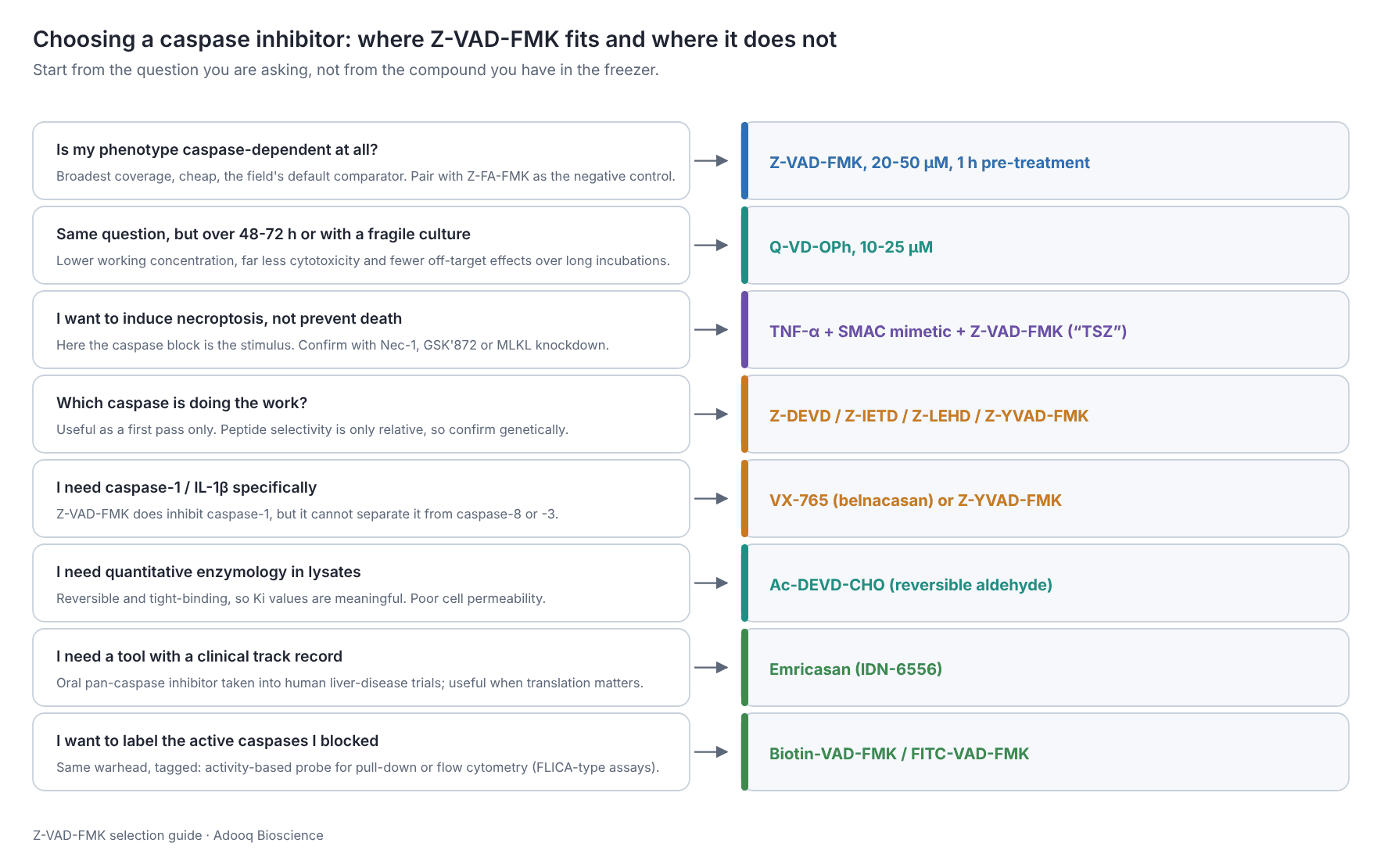
Use Z-VAD-FMK for default screening, broad coverage, and comparability with the existing literature. Switch to Q-VD-OPh for long incubations, fragile or primary cells, in vivo and CNS work, and anywhere inhibitor toxicity would confound the result. Use the selective FMKs for pathway dissection, with genetic confirmation. Use Ac-DEVD-CHO for enzymology, VX-765 for inflammasome-specific claims, and emricasan when translational framing matters.
The strongest papers use two chemically unrelated inhibitors, or one inhibitor plus a genetic manipulation. No single caspase inhibitor is clean enough to carry a conclusion by itself.
9. Troubleshooting
| Symptom | Likely cause | Fix |
|---|---|---|
| No rescue at all | Death is not caspase-dependent, or it switched to necroptosis | Co-treat with Nec-1 and GSK-872; blot p-MLKL |
| Partial rescue only | Concentration too low, inhibitor consumed, or a parallel pathway | Titrate to 50-100 µM; re-dose at 24 h; check PARP cleavage |
| Rescue fades after 24-48 h | Irreversible inhibitor exhausted; new caspase synthesised | Re-dose every 24 h |
| Z-VAD-FMK alone kills cells | Autophagic or necroptotic death (classically L929) or off-target toxicity | Lower the concentration; switch to Q-VD-OPh |
| Works in one line, not another | Esterase activity, RIPK3 expression, caspase pool size | Confirm the block biochemically in each line |
| Precipitate in medium | Aqueous insolubility; stock too dilute | Make a more concentrated DMSO stock; add to warm medium with mixing |
| Inconsistent between batches of stock | Freeze-thaw degradation of the FMK | Single-use aliquots only |
| Phenotype reproduced by Z-FA-FMK | Cathepsin off-target effect | Reinterpret; use Q-VD-OPh |
10. Frequently asked questions
What is the molecular weight of Z-VAD-FMK?
467.49 g/mol for the standard methyl-ester form, Z-VAD(OMe)-FMK (C22H30FN3O7, CAS 187389-52-2). The free acid, Z-VAD(OH)-FMK, is 453.46 g/mol (C21H28FN3O7, CAS 161401-82-7). Check which form you have before calculating molarity.
What is the usual Z-VAD-FMK working concentration?
20-50 µM in cell culture, added 30-60 min before the death stimulus. The full published range is roughly 10-100 µM. Above 100 µM, off-target cysteine protease inhibition and non-specific toxicity become serious confounders.
Is Z-VAD-FMK reversible?
No. It is an irreversible, covalent inhibitor: the fluoromethyl ketone alkylates the caspase catalytic cysteine to form a stable thioether. Washing it out does not restore activity, and recovery requires new caspase synthesis.
What is the difference between Z-VAD-FMK and Z-VAD(OMe)-FMK?
In practice, none. Virtually all commercial Z-VAD-FMK is the O-methylated form. The methyl ester masks the aspartate carboxylate so the molecule can cross the plasma membrane, and intracellular esterases then remove it. Use the OMe form for cells, and the free acid Z-VAD(OH)-FMK for cell-free assays.
Which caspases does Z-VAD-FMK inhibit?
Caspase-1, -3, -4, -5, -6, -7, -8, -9, -10 and murine caspase-11. Caspase-2 is inhibited poorly, and potency is uneven across the family.
Does Z-VAD-FMK block necroptosis?
No. It commonly causes it. By removing caspase-8, which normally cleaves and inactivates RIPK1 and CYLD, Z-VAD-FMK licenses RIPK1-RIPK3 necrosome formation and MLKL-driven lysis. Use Necrostatin-1 or GSK-872 to block necroptosis.
Does Z-VAD-FMK inhibit pyroptosis?
Partly. It inhibits caspase-1 and so suppresses gasdermin D cleavage and IL-1β maturation, but it is not caspase-1-selective. For caspase-1-specific conclusions, use VX-765.
How should I store Z-VAD-FMK?
Lyophilised powder at -20 °C, desiccated and protected from light. Dissolve in anhydrous DMSO, aliquot for single use, store at -20 °C (-80 °C long term) and use within about a month. Avoid repeated freeze-thaw cycles.
Is Z-VAD-FMK soluble in water?
No. It is insoluble in water and ethanol. Use DMSO (80 mg/mL, 171 mM) and dilute into medium immediately before use, keeping final DMSO at or below 0.1%.
What is the best negative control for Z-VAD-FMK?
Z-FA-FMK. It shares the Z-cap and fluoromethyl ketone but lacks the P1 aspartate, so it does not inhibit caspases, while still inhibiting cathepsins. That makes it a direct test for the main off-target liability.
Z-VAD-FMK vs Q-VD-OPh: which should I use?
Z-VAD-FMK for matching the existing literature and for short, standard experiments. Q-VD-OPh when you need activity at lower concentrations, lower cytotoxicity over 48-72 h, brain penetration, or in vivo work.
Can Z-VAD-FMK be used in vivo?
Yes, in rodent models, typically intraperitoneally in a DMSO, PEG 300 and Tween 80 vehicle. Dosing is highly model-dependent. Poor blood-brain-barrier penetration and the FMK fluoroacetate concern are the usual reasons groups switch to Q-VD-OPh or emricasan for animal studies.
11. References
The compound and its chemistry
- Slee EA, Zhu H, Chow SC, MacFarlane M, Nicholson DW, Cohen GM. Benzyloxycarbonyl-Val-Ala-Asp (OMe) fluoromethylketone (Z-VAD.FMK) inhibits apoptosis by blocking the processing of CPP32. Biochem J. 1996;315(Pt 1):21-24. PMID 8670109 · doi:10.1042/bj3150021
- Thornberry NA, Peterson EP, Zhao JJ, Howard AD, Griffin PR, Chapman KT. Inactivation of interleukin-1β converting enzyme by peptide (acyloxy)methyl ketones. Biochemistry. 1994;33(13):3934-3940. PMID 8142397 · doi:10.1021/bi00179a020
- Thornberry NA, Rano TA, Peterson EP, et al. A combinatorial approach defines specificities of members of the caspase family and granzyme B. J Biol Chem. 1997;272(29):17907-17911. PMID 9218414 · doi:10.1074/jbc.272.29.17907
- Garcia-Calvo M, Peterson EP, Leiting B, Ruel R, Nicholson DW, Thornberry NA. Inhibition of human caspases by peptide-based and macromolecular inhibitors. J Biol Chem. 1998;273(49):32608-32613. PMID 9829999 · doi:10.1074/jbc.273.49.32608
- Van Noorden CJF. The history of Z-VAD-FMK, a tool for understanding the significance of caspase inhibition. Acta Histochem. 2001;103(3):241-251. PMID 11482370 · doi:10.1078/0065-1281-00601
Caspase inhibition unmasks necroptosis
- Vercammen D, Beyaert R, Denecker G, et al. Inhibition of caspases increases the sensitivity of L929 cells to necrosis mediated by tumor necrosis factor. J Exp Med. 1998;187(9):1477-1485. PMID 9565639 · doi:10.1084/jem.187.9.1477
- Holler N, Zaru R, Micheau O, et al. Fas triggers an alternative, caspase-8-independent cell death pathway using the kinase RIP as effector molecule. Nat Immunol. 2000;1(6):489-495. PMID 11101870 · doi:10.1038/82732
- Degterev A, Huang Z, Boyce M, et al. Chemical inhibitor of nonapoptotic cell death with therapeutic potential for ischemic brain injury. Nat Chem Biol. 2005;1(2):112-119. PMID 16408008 · doi:10.1038/nchembio711
- Degterev A, Hitomi J, Germscheid M, et al. Identification of RIP1 kinase as a specific cellular target of necrostatins. Nat Chem Biol. 2008;4(5):313-321. PMID 18408713 · doi:10.1038/nchembio.83
- He S, Wang L, Miao L, et al. Receptor interacting protein kinase-3 determines cellular necrotic response to TNF-α. Cell. 2009;137(6):1100-1111. PMID 19524512 · doi:10.1016/j.cell.2009.05.021
- Zhang DW, Shao J, Lin J, et al. RIP3, an energy metabolism regulator that switches TNF-induced cell death from apoptosis to necrosis. Science. 2009;325(5938):332-336. PMID 19498109 · doi:10.1126/science.1172308
- Tenev T, Bianchi K, Darding M, et al. The Ripoptosome, a signaling platform that assembles in response to genotoxic stress and loss of IAPs. Mol Cell. 2011;43(3):432-448. PMID 21737329 · doi:10.1016/j.molcel.2011.06.006
- Feoktistova M, Geserick P, Kellert B, et al. cIAPs block Ripoptosome formation, a RIP1/caspase-8 containing intracellular cell death complex differentially regulated by cFLIP isoforms. Mol Cell. 2011;43(3):449-463. PMID 21737330 · doi:10.1016/j.molcel.2011.06.011
- Mandal P, Berger SB, Pillay S, et al. RIP3 induces apoptosis independent of pronecrotic kinase activity. Mol Cell. 2014;56(4):481-495. PMID 25459880 · doi:10.1016/j.molcel.2014.10.021
- Kaiser WJ, Sridharan H, Huang C, et al. Toll-like receptor 3-mediated necrosis via TRIF, RIP3, and MLKL. J Biol Chem. 2013;288(43):31268-31279. PMID 24019532 · doi:10.1074/jbc.M113.462341
- Linkermann A, Green DR. Necroptosis. N Engl J Med. 2014;370(5):455-465. PMID 24476434 · doi:10.1056/NEJMra1310050
Autophagy and self-inflicted death in L929 cells
- Yu L, Alva A, Su H, et al. Regulation of an ATG7-beclin 1 program of autophagic cell death by caspase-8. Science. 2004;304(5676):1500-1502. PMID 15131264 · doi:10.1126/science.1096645
- Wu YT, Tan HL, Huang Q, et al. Autophagy plays a protective role during zVAD-induced necrotic cell death. Autophagy. 2008;4(4):457-466. PMID 18253089 · doi:10.4161/auto.5662
- Wu YT, Tan HL, Huang Q, et al. zVAD-induced necroptosis in L929 cells depends on autocrine production of TNFα mediated by the PKC-MAPKs-AP-1 pathway. Cell Death Differ. 2011;18(1):26-37. PMID 20539307 · doi:10.1038/cdd.2010.72
Off-target activity and the limits of peptide selectivity
- Schotte P, Declercq W, Van Huffel S, Vandenabeele P, Beyaert R. Non-specific effects of methyl ketone peptide inhibitors of caspases. FEBS Lett. 1999;442(1):117-121. PMID 9923616 · doi:10.1016/S0014-5793(98)01640-8
- Rozman-Pungerčar J, Kopitar-Jerala N, Bogyo M, et al. Inhibition of papain-like cysteine proteases and legumain by caspase-specific inhibitors: when reaction mechanism is more important than specificity. Cell Death Differ. 2003;10(8):881-888. PMID 12867995 · doi:10.1038/sj.cdd.4401247
- Misaghi S, Pacold ME, Blom D, Ploegh HL, Korbel GA. Using a small molecule inhibitor of peptide:N-glycanase to probe its role in glycoprotein turnover. Chem Biol. 2004;11(12):1677-1687. PMID 15610852 · doi:10.1016/j.chembiol.2004.11.010
- McStay GP, Salvesen GS, Green DR. Overlapping cleavage motif selectivity of caspases: implications for analysis of apoptotic pathways. Cell Death Differ. 2008;15(2):322-331. PMID 17975551 · doi:10.1038/sj.cdd.4402260
- Chauvier D, Ankri S, Charriaut-Marlangue C, Casimir R, Jacotot E. Broad-spectrum caspase inhibitors: from myth to reality? Cell Death Differ. 2007;14(2):387-391. PMID 17008913 · doi:10.1038/sj.cdd.4402044
Alternatives and in vivo work
- Caserta TM, Smith AN, Gultice AD, Reedy MA, Brown TL. Q-VD-OPh, a broad spectrum caspase inhibitor with potent antiapoptotic properties. Apoptosis. 2003;8(4):345-352. PMID 12815277 · doi:10.1023/A:1024116916932
- Rodriguez I, Matsuura K, Ody C, Nagata S, Vassalli P. Systemic injection of a tripeptide inhibits the intracellular activation of CPP32-like proteases in vivo and fully protects mice against Fas-mediated fulminant liver destruction and death. J Exp Med. 1996;184(5):2067-2072. PMID 8920897 · doi:10.1084/jem.184.5.2067
- Hara H, Friedlander RM, Gagliardini V, et al. Inhibition of interleukin 1β converting enzyme family proteases reduces ischemic and excitotoxic neuronal damage. Proc Natl Acad Sci U S A. 1997;94(5):2007-2012. PMID 9050895 · doi:10.1073/pnas.94.5.2007
- Hotchkiss RS, Chang KC, Swanson PE, et al. Caspase inhibitors improve survival in sepsis: a critical role of the lymphocyte. Nat Immunol. 2000;1(6):496-501. PMID 11101871 · doi:10.1038/82741
- Wannamaker W, Davies R, Namchuk M, et al. VX-765, an orally available selective interleukin-converting enzyme/caspase-1 inhibitor, exhibits potent anti-inflammatory activities by inhibiting the release of IL-1β and IL-18. J Pharmacol Exp Ther. 2007;321(2):509-516. PMID 17289835 · doi:10.1124/jpet.106.111344 (title shortened; the indexed title begins with the full chemical name)
- Brumatti G, Ma C, Lalaoui N, et al. The caspase-8 inhibitor emricasan combines with the SMAC mimetic birinapant to induce necroptosis and treat acute myeloid leukemia. Sci Transl Med. 2016;8(339):339ra69. PMID 27194727 · doi:10.1126/scitranslmed.aad3099
- Garcia-Tsao G, Fuchs M, Shiffman M, et al. Emricasan (IDN-6556) lowers portal pressure in patients with compensated cirrhosis and severe portal hypertension. Hepatology. 2019;69(2):717-728. PMID 30063802 · doi:10.1002/hep.30199
- Garcia-Tsao G, Bosch J, Kayali Z, et al. Randomized placebo-controlled trial of emricasan for non-alcoholic steatohepatitis-related cirrhosis with severe portal hypertension. J Hepatol. 2020;72(5):885-895. PMID 31870950 · doi:10.1016/j.jhep.2019.12.010
Pyroptosis
- Fink SL, Cookson BT. Caspase-1-dependent pore formation during pyroptosis leads to osmotic lysis of infected host macrophages. Cell Microbiol. 2006;8(11):1812-1825. PMID 16824040 · doi:10.1111/j.1462-5822.2006.00751.x
Product data
- Adooq Bioscience. Z-VAD-FMK (A12373) product data: CAS 187389-52-2, C22H30FN3O7, MW 467.49, DMSO solubility 80 mg/mL. https://www.adooq.com/z-vad-fmk.html
- Adooq Bioscience. Z-VAD(OH)-FMK (A16340) and Q-VD-OPh hydrate (A14915) product data, including the Q-VD-OPh caspase IC50 values quoted in §8. https://www.adooq.com/z-vad-oh-fmk.html · https://www.adooq.com/q-vd-oph-hydrate.html
- InvivoGen. Z-VAD-FMK technical data sheet (working range 1-10 µg/mL; caspase coverage; caspase-2 exception).
Z-VAD-FMK is supplied for research use only. Not for human or veterinary diagnostic or therapeutic use.