
Necrostatin-1: Mechanism of Action, Working Concentration, and What the Clinical Record Actually Shows
How Necrostatin-1 inhibits RIPK1, what working concentration to use, which controls rule out its IDO off-target effect, and why Nec-1 itself has never entered a clinical trial.
Technical guide from Adooq Bioscience · Product: Necrostatin-1, Cat. No. A11973 · Last reviewed: November 2026
Necrostatin-1 at a glance
| Property | Value |
|---|---|
| Common name | Necrostatin-1 (Nec-1) |
| Other names | MTH-Trp (methyl-thiohydantoin-tryptophan), 5-(1H-indol-3-ylmethyl)-3-methyl-2-thioxo-4-imidazolidinone |
| CAS | 4311-88-0 |
| Molecular formula | C13H13N3OS |
| Necrostatin-1 molecular weight | 259.33 g/mol |
| Target | RIPK1 (RIP1) kinase. ATP-competitive, binds an inactive conformation of the kinase domain |
| Potency | EC50 494 nM against TNF-induced necroptosis in FADD-deficient Jurkat cells; EC50 182 nM against endogenous RIP1 kinase activity |
| Known off-target | Indoleamine 2,3-dioxygenase (IDO), IC50 11.4 µM |
| Solubility | DMSO 43 mg/mL (165.8 mM) with warming; insoluble in water; ethanol and methanol poor (3-5 mg/mL) |
| Typical stock | 10-20 mM in DMSO, single-use aliquots, -20 °C |
| Necrostatin-1 working concentration | 10-50 µM in cell culture; 30 µM is the most common single value, 10-100 µM is the published range |
| Pre-treatment | 30-60 min before the necroptosis stimulus |
| Metabolic stability | T1/2 under 5 min in mouse liver microsomes. This is the single most important fact about using it in animals |
| In vivo vehicle | 5% DMSO / 45% PEG 300 / ddH2O, to about 9 mg/mL |
| Storage | -20 °C desiccated, protected from light; DMSO stocks for about a month at -20 °C |
| Clinical trials | None. Necrostatin-1 has never been tested in humans. More than ten other RIPK1 inhibitors have (see §8) |
Necrostatin-1 is the compound that made necroptosis a tractable field. Twenty years on it is also the least specific and least stable reagent in that field, and its two off-target liabilities are documented in unusual detail, including in an addendum its own discoverers published. Both facts shape how the compound should be used.
1. What Necrostatin-1 is
1.1 Origin
Necrostatin-1 came out of a phenotypic screen, not a target-based campaign. Degterev and colleagues treated human U937 cells with TNF-α plus a caspase inhibitor, which kills them by a non-apoptotic route, and screened roughly 15,000 compounds for protection. The hit they called necrostatin-1 blocked that death without blocking apoptosis. In the same paper they named the pathway necroptosis and showed that it contributes to delayed ischaemic brain injury in mice (Degterev et al. 2005).
The target came three years later. Nec-1 inhibits RIPK1 kinase activity, it is ATP-competitive, and it has no effect on RIPK2 or RIPK3 autophosphorylation (Degterev et al. 2008). That sequence matters when you read the literature. Papers from 2005 to 2008 describe the same compound as a "necroptosis inhibitor" of unknown mechanism, and papers after 2008 describe it as a RIPK1 inhibitor.
1.2 Structure
Nec-1 is a thiohydantoin bearing an indol-3-ylmethyl group, which is to say a cyclised, methylated tryptophan derivative. Two parts of that structure cause most of the trouble, and a third is often overlooked.
The 2-thioxo sulfur is why the molecule is metabolically fragile. Degterev and colleagues measured a half-life of under 5 minutes in a mouse microsomal assay and attributed it to that sulfur (Degterev et al. 2013b). The optimised analogue in which the sulfur is replaced by oxygen has a microsomal half-life of about 1 hour.
The indole is the off-target problem. Nec-1 is chemically identical to methyl-thiohydantoin-tryptophan, MTH-Trp, which was already in the literature as an inhibitor of indoleamine 2,3-dioxygenase. The same compound had been sold under two names for different purposes (Vandenabeele et al. 2013). The tryptophan-like indole is what IDO recognises, so the off-target is structural and cannot be dialled out by careful pipetting.
The third point is stereochemistry. Commercial Nec-1 is the racemate. In the chlorinated hydantoin series the (R)-enantiomer is about four-fold more potent than the (S)-enantiomer (EC50 50 nM versus 230 nM), so roughly half of what you weigh out is the weaker isomer.
1.3 Nec-1, Nec-1s, Nec-1i and Nec-2: which vial is which
The names are inconsistent across catalogues and the compounds are not interchangeable, which is where most mix-ups in this field start.
| Name in papers | Also sold as | CAS | Formula / MW | What it is |
|---|---|---|---|---|
| Nec-1 | Necrostatin-1, MTH-Trp | 4311-88-0 | C13H13N3OS / 259.33 | The original. Racemic thiohydantoin. Inhibits RIPK1 and IDO. Microsomal T1/2 < 5 min |
| Nec-1s, 7-Cl-O-Nec-1 | "Necrostatin 2 racemate", (±)-Nec-2 | 852391-15-2 | C13H12ClN3O2 / 277.71 | Sulfur replaced by oxygen, chlorine added to the indole. Equipotent on RIPK1, no IDO activity, T1/2 about 1 h, selective among more than 400 human kinases |
| Nec-2 | Necrostatin-2 | 852391-19-6 | C13H12ClN3O2 / 277.71 | The (5R)-enantiomer of the above. EC50 50 nM; the (S)-enantiomer is 230 nM |
| Nec-1i | Necrostatin-1 inactive control | n/a | demethylated Nec-1 | Sold as an inactive control. It is not one in vivo (Takahashi et al. 2012) |
Two practical consequences. First, if a paper says "Nec-1s" and you want to reproduce it, the vial you need may be labelled necrostatin-2 racemate; at Adooq that is A15327, CAS 852391-15-2. Second, Necrostatin 2 (A15183, CAS 852391-19-6) is the single enantiomer of that same optimised scaffold, not a different generation of inhibitor.
2. Necrostatin-1 mechanism of action
2.1 What RIPK1 does, and why blocking its kinase activity is not the same as removing it
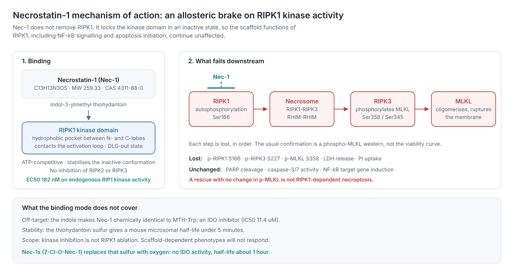
RIPK1 sits immediately downstream of TNFR1 and has two separable jobs. As a scaffold, through its intermediate domain and death domain, it supports NF-κB survival signalling and the assembly of complex I. As a kinase, through autophosphorylation at Ser166 and recruitment of RIPK3 through the RHIM domains, it drives necrosome formation. Nec-1 only touches the second job.
The order of events that Nec-1 interrupts:
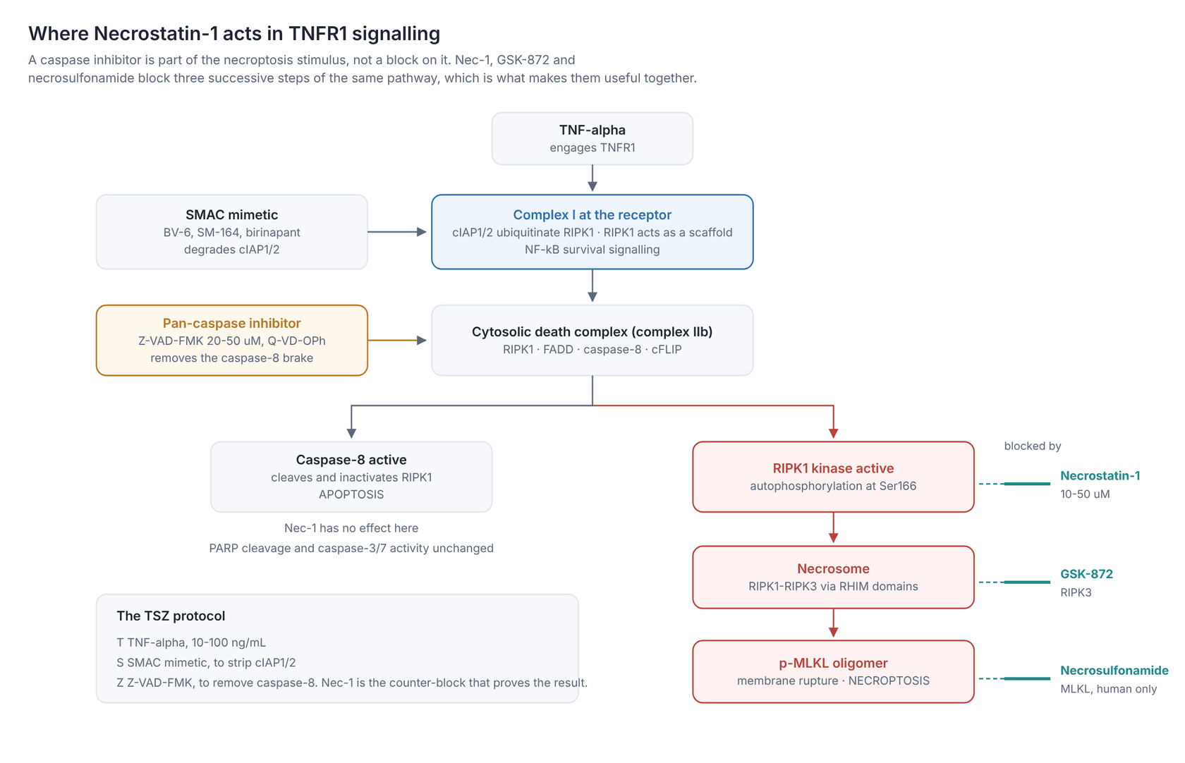
- TNFR1 ligation builds complex I. cIAP1/2-mediated ubiquitination of RIPK1 keeps it in scaffold mode.
- If cIAPs are depleted, by a SMAC mimetic or genetically, RIPK1 is deubiquitinated and moves into the cytosolic death complex.
- Caspase-8, with FADD and cFLIP, normally cleaves RIPK1 and shuts the pathway down. Block caspase-8 (a caspase inhibitor, or FADD deficiency) and that brake is gone.
- RIPK1 autophosphorylates at Ser166, engages RIPK3 through RHIM-RHIM interaction, and the necrosome forms.
- RIPK3 phosphorylates MLKL at Ser358 (human) or Ser345 (mouse). Phospho-MLKL oligomerises, moves to the plasma membrane, and ruptures it.
Nec-1 blocks step 4. Everything downstream fails: no RIPK1 Ser166 phosphorylation, no RIPK3 Ser227 phosphorylation, no phospho-MLKL, no lysis, no LDH release. Everything upstream is untouched, which is why Nec-1 does not rescue apoptosis and does not restore NF-κB signalling.
2.2 How Nec-1 binds the kinase
Degterev and colleagues showed the inhibition is ATP-competitive and modelled Nec-1 into the ATP cleft, predicting hydrogen bonds to the hinge residues Glu93, Tyr94, Glu96, Lys97 and Asn99 and hydrophobic contacts with Leu23, Leu143, Met92 and Leu157, with the kinase in its inactive DLG-out state (Degterev et al. 2008).
The crystal structures settled it. Xie and colleagues solved the RIPK1 kinase domain with necrostatins bound and found the inhibitor caged in a hydrophobic pocket between the N- and C-lobes, making contacts with conserved activation-loop residues that lock the kinase in an inactive conformation (Xie et al. 2013; PDB 4ITH, 4ITI, 4ITJ). Necrostatins are therefore best described as type III-like allosteric inhibitors of an inactive state, which is also the structural reason they are selective: the pocket they use is not conserved across the kinome the way the ATP site is.
2.3 The potency numbers, and which one to quote
Three numbers circulate, and they measure different things:
- EC50 494 nM: protection of FADD-deficient Jurkat cells from 30 h of TNF-α (10 ng/mL). This is a cellular, phenotypic number. Some catalogues round it to 490 nM.
- EC50 182 nM: inhibition of endogenous RIP1 kinase activity recovered from Jurkat cells (Degterev et al. 2008). This is the biochemical number, and the one on the Adooq datasheet.
- IC50 11.4 µM: inhibition of IDO. This is the off-target number, and it is the one that constrains your working concentration (§7.1).
The gap between a 494 nM cellular EC50 and a 10-50 µM working concentration is wide. Cellular potency in a 30 h Jurkat assay does not transfer to a 6 h assay in primary cells: Nec-1 degrades quickly, dissolves poorly in aqueous medium, and competes against a kinase that is being actively recruited. Titrate in your own system instead of reasoning down from the EC50.
2.4 Readouts that confirm the block is real
| Readout | Expected with Nec-1 |
|---|---|
| Phospho-RIPK1 Ser166 (western) | Lost. The most direct target-engagement readout available |
| Phospho-RIPK3 Ser227 (human) / Ser232 (mouse) | Lost |
| Phospho-MLKL Ser358 (human) / Ser345 (mouse) | Lost. The standard necroptosis marker |
| RIPK1-RIPK3 co-immunoprecipitation | Reduced |
| LDH release, PI uptake, SYTOX Green | Reduced |
| PARP cleavage, caspase-3/7 activity | Unchanged. Nec-1 does not block apoptosis |
| NF-κB target gene induction | Unchanged. The scaffold function is intact |
If the viability curve moves but phospho-MLKL does not, the rescue is not RIPK1-dependent necroptosis. That pattern is the main way the IDO and RIPK1-independent effects discussed in §7 get caught.
3. Why researchers use Necrostatin-1: four distinct jobs
The right concentration, timing and controls differ depending on which of these experiments you are actually running.
Testing whether a death is necroptotic is the default use. A stimulus kills cells, Nec-1 prevents it, and the death is therefore RIPK1-kinase-dependent. This is the backbone of the field, and it is also where the compound's specificity problems bite hardest, because the conclusion rests entirely on the inhibitor being clean.
In pathway triage, Nec-1 is one arm of the standard four-inhibitor classification of an unknown death, alongside a pan-caspase inhibitor, a ferroptosis inhibitor and an autophagy blocker (§4.5).
In injury models, stroke, spinal cord injury, ischaemia-reperfusion, TBI and sepsis models all use Nec-1 to ask whether necroptosis contributes to the lesion. This is the largest body of Nec-1 literature and also the body most exposed to the compound's half-life problem.
Pairing Nec-1 with a caspase inhibitor in the same experiment separates "death requires caspases" from "death requires RIPK1 kinase activity" from "the cell switches between the two". A death that is blocked only by both compounds together is the signature of a system that reroutes.
4. Experiment types and research areas
4.1 Necroptosis in cell culture
The canonical induction cocktail is TNF-α plus a SMAC mimetic plus a pan-caspase inhibitor, abbreviated TSZ. Nec-1 is the counter-block that proves the death was RIPK1-dependent.
| Component | Typical reagent | Role |
|---|---|---|
| T | TNF-α, 10-100 ng/mL | Engages TNFR1 |
| S | BV-6, SM-164 or birinapant | Degrades cIAP1/2, releases RIPK1 from scaffold mode |
| Z | Z-VAD-FMK 20-50 µM, or Q-VD-OPh | Removes caspase-8, the brake on RIPK1 |
| Counter-block | Necrostatin-1, 10-50 µM | Should abolish the death |
Cell lines that run this reliably: human HT-29 and HeLa (with RIPK3 restored), human U937 and Jurkat (FADD-deficient Jurkat is the historical assay), mouse L929 (which needs only TNF plus a caspase inhibitor), mouse MEFs, and bone-marrow-derived macrophages. RIPK3 expression is the gatekeeper; many common lines, including MCF-7 and most long-passaged cancer lines, have silenced RIPK3 and cannot do necroptosis at all, so a negative Nec-1 result in them means nothing.
4.2 Stroke, ischaemia-reperfusion and the CNS
This is where the compound started. The 2005 paper showed dose-dependent infarct reduction in a mouse model of cerebral ischaemia, and, unusually for a neuroprotectant, protection when administration was delayed by up to 6 hours after the insult, which is longer than the window for caspase inhibitors (Degterev et al. 2005). That window is a large part of why the pathway attracted translational interest.
The same logic has since been applied to myocardial ischaemia-reperfusion, renal IRI, hepatic IRI, spinal cord injury, traumatic brain injury, subarachnoid haemorrhage and retinal degeneration. Necroptosis is also activated in human multiple sclerosis cortical lesions, where RIPK1 and RIPK3 are found in detergent-insoluble complexes and RIPK1 carries Ser14/15 and Ser166 phosphorylation (Ofengeim et al. 2015).
Before designing CNS dosing, see §5.5: the half-life is short, and the original brain work delivered the compound intracerebroventricularly rather than systemically.
4.3 Inflammation, sepsis and systemic inflammatory response
TNF-induced systemic inflammatory response syndrome (SIRS) in mice is the standard in vivo necroptosis model, and it is also the model that exposed Nec-1's dose paradox. Takahashi and colleagues found that 3 and 6 mg/kg i.v. protected mice against an otherwise lethal TNF challenge, while 0.6 mg/kg of Nec-1 sensitised them, producing faster hypothermia and lower survival. Nec-1s did not do this at any dose tested (Takahashi et al. 2012). If your in vivo result is a worsening rather than a rescue, underdosing is a live explanation.
4.4 Infection, immunity and cancer biology
Viral and bacterial infection models use Nec-1 to separate caspase-dependent death of infected cells from RIPK3-driven necroptosis, which many pathogens actively suppress. In tumour biology the interest runs both ways: necroptosis as an immunogenic cell death to be induced, and RIPK1 signalling as a driver of the inflammatory tumour microenvironment to be blocked. Nec-1 appears on both sides.
4.5 Cell-death-pathway triage
| Add | If death is blocked, suspect |
|---|---|
| Z-VAD-FMK (20-50 µM) | Apoptosis or pyroptosis |
| Necrostatin-1 (10-50 µM) | Necroptosis, RIPK1-kinase-dependent |
| Ferrostatin-1 or liproxstatin-1 | Ferroptosis |
| 3-Methyladenine or bafilomycin A1 | Autophagy-dependent death |
One caveat specific to this panel: Nec-1 itself blocks ferroptosis in some cell lines through a mechanism independent of both RIPK1 and IDO, so the two arms of this panel are not cleanly separable with Nec-1 alone (§7.3).
5. Necrostatin-1 working concentration: a practical protocol
5.1 Stock solution
- Spin the vial before opening.
- Dissolve in DMSO. Adooq reports 43 mg/mL (165.8 mM) with warming; other suppliers list 10-20 mg/mL for the same compound, so warm to 37 °C and vortex rather than assuming a failed dissolution. A 10 mM or 20 mM stock is a comfortable working choice (1 mg of MW 259.33 material in 193 µL gives 20 mM).
- The compound is insoluble in water and poorly soluble in ethanol and methanol (3-5 mg/mL). Do not try to make an aqueous stock.
- Aliquot single-use volumes and store at -20 °C. Use DMSO stocks within about a month; the thiohydantoin is the reactive part of the molecule and does not improve with freeze-thaw cycles.
- Keep final DMSO at or below 0.1% (v/v) and run a vehicle-matched control. At 50 µM from a 10 mM stock, DMSO is 0.5%, which is too high for many primary cells; use a 20 mM stock instead.
- Dilute into pre-warmed medium immediately before use, with mixing. Nec-1 precipitates out of cold medium.
5.2 Choosing the concentration
| Application | Necrostatin-1 concentration | Timing | Notes |
|---|---|---|---|
| Standard necroptosis block (most cell lines) | 10-50 µM, start at 30 µM | 30-60 min pre-treatment, then co-incubate | 30 µM is the field's default single value |
| L929, HT-29, U937: well-characterised lines | 10-30 µM | 30 min pre-treatment | These lines are sensitive; higher adds off-target risk |
| Primary cells, slow uptake | 30-50 µM | 60 min pre-treatment | Titrate; confirm with phospho-MLKL |
| Resistant systems | up to 100 µM | 1 h pre-treatment, re-dose at 12-24 h | Above about 30 µM you are also inhibiting IDO (§7.1); above 50 µM, interpret with a Nec-1s arm |
| Long experiments (over 24 h) | 10-50 µM, re-dosed every 12-24 h | n/a | Short half-life, not inhibitor consumption, is the issue |
| Biochemical RIPK1 kinase assay | 0.1-10 µM | 15-30 min pre-incubation | The EC50 is sub-micromolar here; µM-range cell doses are not needed in vitro |
| In vivo (rodent) | See §5.5. Prefer Nec-1s | n/a | Formulate at up to about 9 mg/mL in 5% DMSO / 45% PEG 300 / water |
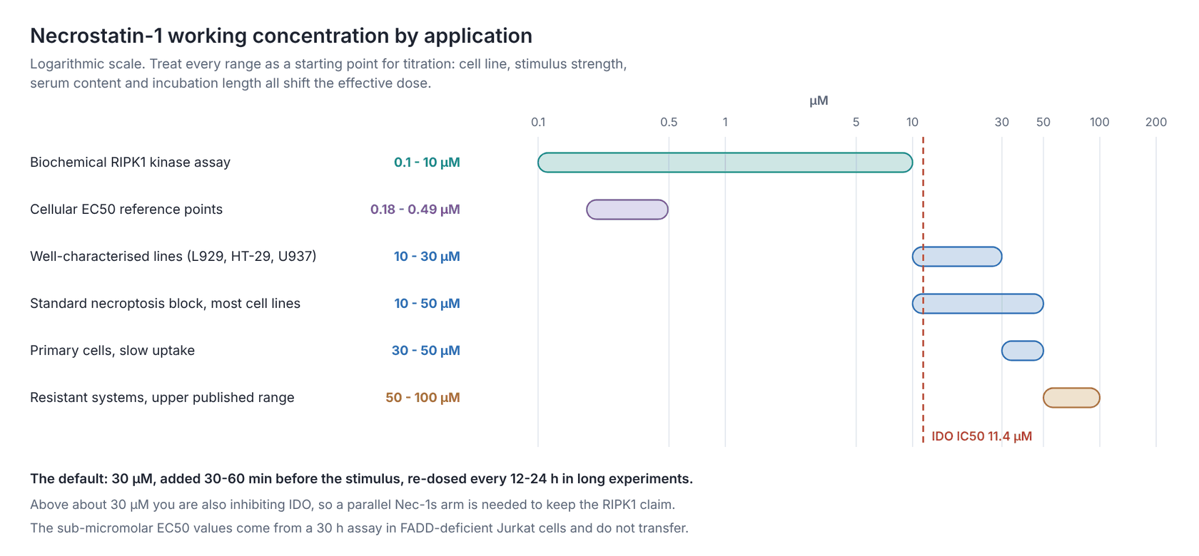
Above roughly 30 µM you are above the IDO IC50 of 11.4 µM by more than two-fold, so an IDO-dependent phenotype becomes plausible. The necroptosis EC50 and the IDO IC50 are separated by only about 20-fold (Degterev et al. 2013a), which is narrow by the standards of chemical biology, and it is what sets the practical upper bound.
5.3 Timing rules that matter
- Pre-incubate for 30-60 min. Nec-1 has to be bound before RIPK1 is recruited and autophosphorylated. Adding it with the stimulus loses part of the effect; adding it after the necrosome has formed loses most of it.
- Re-dose anything longer than a day. With a microsomal half-life under 5 minutes, and limited stability in serum-containing medium, a single dose at t = 0 does not cover a 48 h experiment. Re-dosing every 12-24 h is the practical fix; switching to Nec-1s is the better one.
- Washout reversal tells you little. Nec-1 is reversible, so washing it out does restore RIPK1 activity, but the necrosome is a positive-feedback assembly, and timing dominates. Use an add-back time course instead.
5.4 Controls you should run
| Control | Purpose |
|---|---|
| DMSO vehicle, concentration-matched | Rules out solvent effects, which matter here because the required DMSO can reach 0.5% |
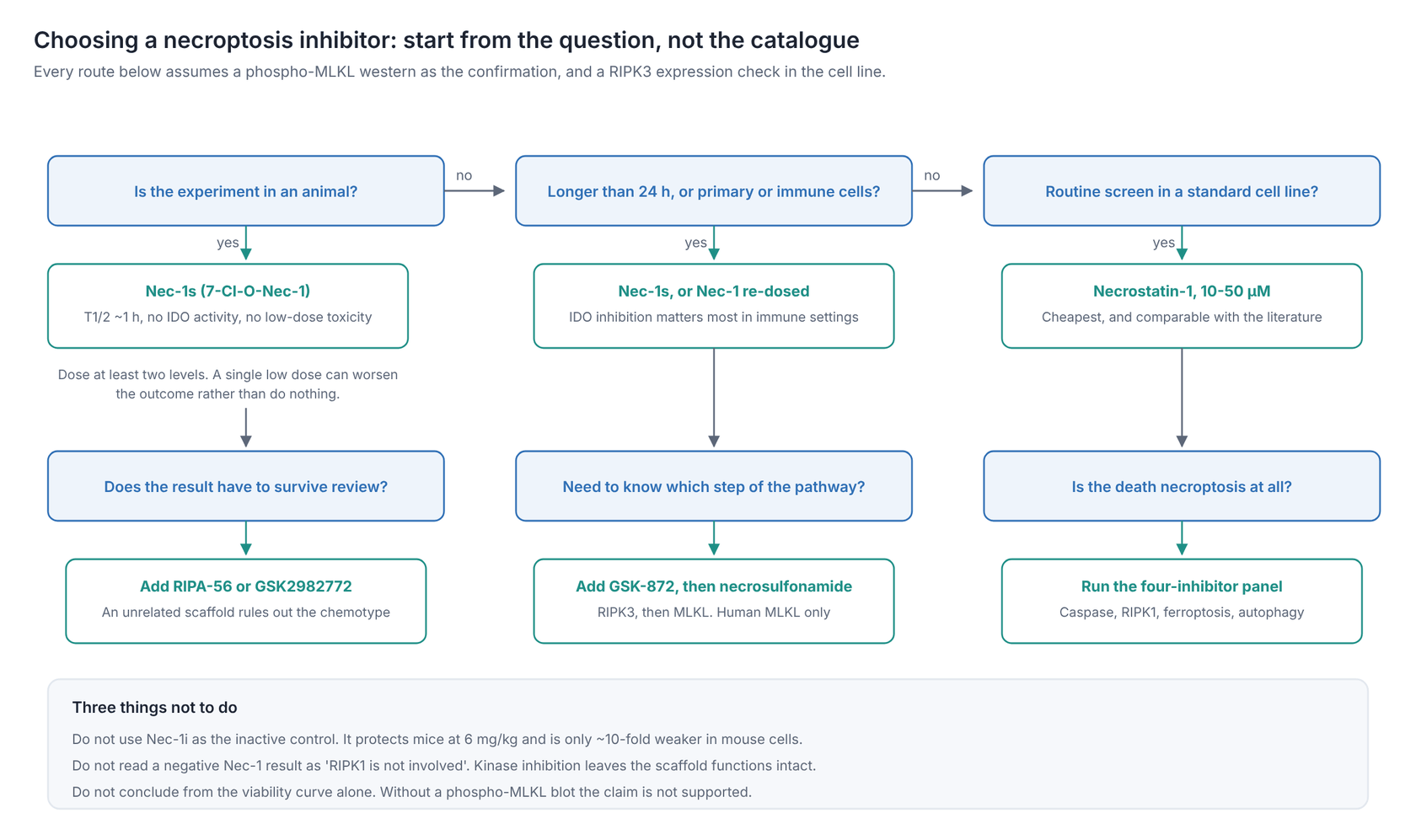
| Nec-1s / 7-Cl-O-Nec-1 in parallel | The most useful single control. Same target, no IDO activity, better stability. A phenotype reproduced by Nec-1s is RIPK1-dependent; one that is not is probably not |
| An IDO inhibitor such as 1-methyl-tryptophan | Tests the IDO off-target directly. 1-MT does not inhibit necroptosis (Degterev et al. 2013b), so if 1-MT reproduces your phenotype, the phenotype is IDO |
| GSK-872 (RIPK3) or necrosulfonamide (MLKL, human) | Block the step below RIPK1. Agreement across three pathway nodes is far stronger than any single inhibitor |
| A structurally unrelated RIPK1 inhibitor, e.g. RIPA-56 or GSK2982772 | Rules out scaffold-specific off-targets entirely |
| Phospho-MLKL western | Confirms the block is real at the molecular level, not just on the viability curve |
| RIPK3 expression check in your line | A line without RIPK3 cannot undergo necroptosis; a Nec-1 effect there is off-target by definition |
| RIPK1 knockdown, knockout, or kinase-dead knock-in | The genetic confirmation that makes the pharmacology conclusive |
Do not use Nec-1i as your inactive control. It is sold for that purpose, but Takahashi and colleagues showed it does not qualify: it is more than 100-fold weaker than Nec-1 on human RIPK1 in vitro but only about 10-fold weaker in mouse L929sA necroptosis assays, becoming equipotent at 10-100 µM, and at 6 mg/kg i.v. it protected mice against TNF-induced SIRS as well as Nec-1 did (Takahashi et al. 2012). An "inactive control" that is active at your working concentration is worse than no control.
5.5 In vivo use
Necrostatin-1 is used in animals widely and successfully. It is still the wrong default choice for a new study, for pharmacokinetic reasons.
Four facts govern the design:
- The microsomal half-life is under 5 minutes in mouse liver microsomes (Degterev et al. 2013b). The optimised analogue Nec-1s is roughly 1 hour.
- Nec-1s is the compound its discoverers recommend. The 2013 addendum to the original 2005 paper recommends 7-Cl-O-Nec-1 for both in-cell and in vivo work, on the grounds of higher activity, better metabolic stability, no IDO activity, and no paradoxical toxicity (Degterev et al. 2013a).
- Low doses can worsen the outcome: 0.6 mg/kg Nec-1 sensitised mice to TNF lethality while 3 and 6 mg/kg protected (Takahashi et al. 2012).
- CNS delivery is the historical route. Published murine MCAO work with the 7-chloro analogue used intracerebroventricular dosing, two injections of 8 nmol in 2 µL per mouse, rather than relying on systemic exposure.
Practical points:
- Take the dose, route and schedule from the paper closest to your model. Published Nec-1 regimens span intraperitoneal, intravenous, intracerebroventricular, intrathecal and intranasal routes across stroke, SCI, SAH, TBI, renal and cardiac IRI, epilepsy and EAE models, and the doses differ by more than an order of magnitude between them. A dose transplanted from an unrelated model is a guess.
- Formulate fresh, for example at up to about 9 mg/mL in 5% DMSO / 45% PEG 300 / water.
- Include a dose above and below your target. Given the sensitisation finding, a single-dose in vivo experiment cannot distinguish "no effect" from "underdosed into the harmful range".
- Consider switching to Nec-1s before you start. For a new in vivo programme there is no literature argument for Nec-1 over Nec-1s, only a cost one.
6. Molecules commonly used alongside Necrostatin-1
| Molecule | Role | Why it appears with Necrostatin-1 |
|---|---|---|
| TNF-α | Death-receptor ligand | The standard necroptosis trigger |
| Z-VAD-FMK, Q-VD-OPh | Pan-caspase inhibitors | Remove caspase-8, which is what permits necroptosis in the first place |
| BV-6, SM-164, birinapant | SMAC mimetics / IAP antagonists | Degrade cIAP1/2 so RIPK1 leaves scaffold mode |
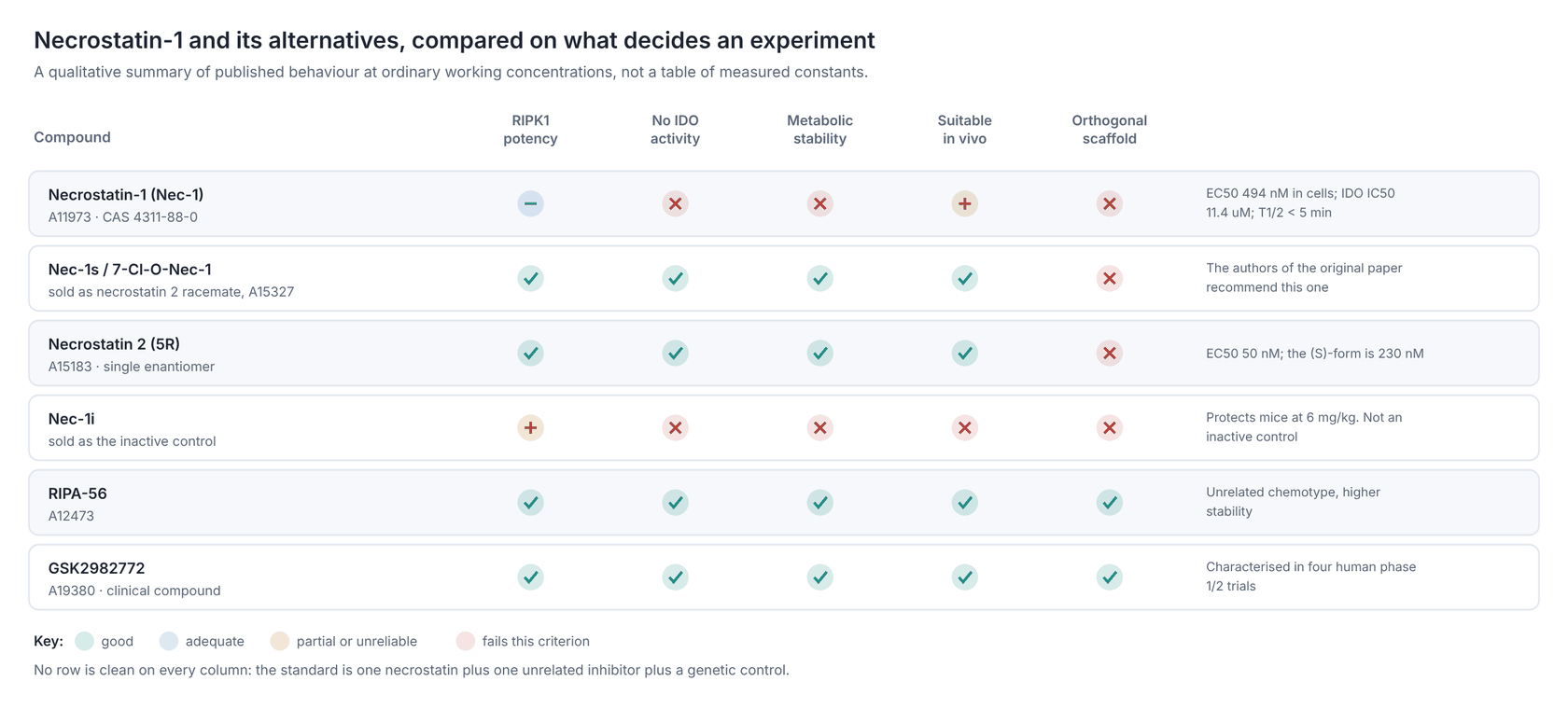
| Nec-1s (7-Cl-O-Nec-1), Necrostatin 2 | Optimised RIPK1 inhibitors | The specificity and stability controls for every Nec-1 experiment |
| RIPA-56, GSK2982772 | Chemically unrelated RIPK1 inhibitors | Confirm the target independently of the necrostatin scaffold |
| GSK-872, GSK-843 | RIPK3 inhibitors | Block the step below RIPK1 |
| Necrosulfonamide | MLKL inhibitor (human) | Blocks the terminal step |
| Staurosporine | Apoptosis positive control | Nec-1 should not rescue it |
| Ferrostatin-1, liproxstatin-1 | Ferroptosis inhibitors | Pathway triage, and the control for Nec-1's own anti-ferroptotic activity |
| 3-Methyladenine, bafilomycin A1 | Autophagy inhibitors | Pathway triage |
| 1-methyl-tryptophan | IDO inhibitor | The direct control for Nec-1's main off-target |
7. Limitations and off-target effects
Necrostatin-1 is a first-in-class tool compound from 2005 with two documented off-target activities, poor metabolic stability, a commercial "inactive control" that is not inactive, and a dose-response that can invert in vivo. Three of those five problems were published by the group that discovered the compound.
7.1 It inhibits IDO
Nec-1 is chemically identical to MTH-Trp, a known inhibitor of indoleamine 2,3-dioxygenase, and inhibits IDO with an IC50 of 11.4 µM. IDO controls tryptophan catabolism along the kynurenine pathway, with well-documented effects on T-cell function, tumour immune evasion and neuroinflammation, so the off-target is biologically active in exactly the settings where Nec-1 is most used (Vandenabeele et al. 2013; Takahashi et al. 2012).
The compound is only about 20-fold more potent against necroptosis than against IDO (Degterev et al. 2013a), so at a 30-50 µM working concentration you are inhibiting both. To control for it, run Nec-1s, which has no IDO activity, alongside 1-methyl-tryptophan (1-MT), which inhibits IDO and does not inhibit necroptosis.
7.2 Nec-1i is not an inactive control
Covered in §5.4. The compound sold as the inactive analogue protects mice against TNF-induced SIRS as effectively as Nec-1 at 6 mg/kg, and is only about 10-fold weaker in mouse necroptosis assays (Takahashi et al. 2012). Published experiments that rest on Nec-1i as their specificity control are not controlled.
7.3 It blocks ferroptosis independently of RIPK1 and IDO
In Huh7 and SK-HEP-1 hepatocellular carcinoma cells, 20 µM Nec-1 prevented sulfasalazine-induced ferroptosis, while Nec-1s, GSK2982772 and 1-MT did not, which places the effect outside both RIPK1 and IDO (Yuk et al. 2021). The authors attribute it to the structure of Nec-1 itself. For the triage panel in §4.5 that means a death blocked by both Nec-1 and ferrostatin-1 is not necessarily two pathways.
7.4 Metabolic instability
A microsomal half-life under 5 minutes (Degterev et al. 2013b) means in vivo exposure is brief and highly variable between protocols, and that long in vitro incubations lose inhibitor over time. A "Nec-1 stopped working at 48 h" result is usually this and not biology.
7.5 The dose-response can invert
0.6 mg/kg sensitised mice to TNF lethality; 3 and 6 mg/kg protected (Takahashi et al. 2012). The authors suggest this may explain part of the conflicting literature on RIPK1 in SIRS and sepsis. Any in vivo Nec-1 experiment needs more than one dose.
7.6 Kinase-activity inhibition is not RIPK1 ablation
Nec-1 blocks the kinase function and leaves the scaffold function intact. Phenotypes that depend on RIPK1 as a scaffold, including much of its role in NF-κB signalling and in apoptosis initiation, will not respond to Nec-1 at all. A negative Nec-1 result does not exclude RIPK1 involvement; it excludes RIPK1 kinase involvement.
7.7 Racemic material
Commercial Nec-1 is a racemate, and in the closely related chlorinated series the enantiomers differ about four-fold in potency. Batch-to-batch enantiomeric composition is not normally specified.
8. Necrostatin-1 clinical trials: the honest answer
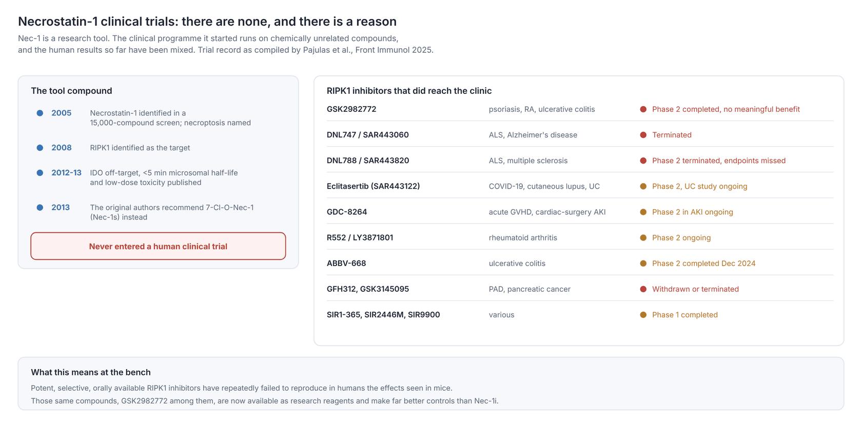
Necrostatin-1 has never been tested in a human clinical trial. It is a research tool, and the properties documented in §7 are each on their own disqualifying for drug development: a microsomal half-life under 5 minutes, an IDO off-target at 11.4 µM, a racemic structure, and a dose-response that inverts in mice. It appears nowhere in the compiled registry record of RIPK1 inhibitor trials (Pajulas et al. 2025).
What exists instead is a substantial clinical programme built on the target Nec-1 identified. RIPK1 inhibitors from at least eight companies have entered human trials. The table below is drawn from a 2025 review that compiled the registry record (Pajulas et al. 2025).
| Compound (sponsor) | Highest phase | Indications tested | Status and outcome |
|---|---|---|---|
| GSK2982772 (GSK) | Phase 2 | Plaque psoriasis, rheumatoid arthritis, ulcerative colitis | Completed. Reduced epidermal thickness and T-cell infiltration in mild-moderate psoriasis (NCT02776033). No meaningful benefit over placebo in RA (NCT02858492, Weisel et al. 2021) or UC (NCT02903966). In chronic plaque psoriasis, PASI75 at week 12 was reached by 1 of 19 patients on drug versus 0 of 10 on placebo, and the authors concluded that target inhibition "did not translate into meaningful clinical improvements" (Ludbrook et al. 2024) |
| DNL747 / SAR443060 (Denali, Sanofi) | Phase 1 | ALS, Alzheimer's disease | Terminated. The ALS study stopped on a strategy change; the AD study completed but development was discontinued over dose-limiting toxicity risk |
| Oditrasertib (DNL788 / SAR443820, Denali and Sanofi) | Phase 2 | ALS, multiple sclerosis | Terminated. Brain-penetrant, but the ALS study (NCT05237284) missed its primary endpoint and the MS study (NCT05630547) missed primary and key secondary endpoints |
| Eclitasertib (DNL758 / SAR443122, Denali and Sanofi) | Phase 2 | COVID-19, cutaneous lupus, ulcerative colitis | Peripherally restricted. COVID-19 and lupus studies completed; the UC study (NCT05588843) was listed as recruiting |
| Flizasertib (GDC-8264, Genentech) | Phase 2 | Acute GVHD, cardiac-surgery-associated AKI | The aGVHD study was terminated for business reasons; the AKI study (NCT06602453) was listed as recruiting |
| Ocadusertib (R552 / LY3871801, Rigel and Eli Lilly) | Phase 2 | Rheumatoid arthritis | Phase 1 completed with a prolonged half-life and good tolerability; the RA study (NCT05848258) was listed as recruiting |
| ABBV-668 (AbbVie) | Phase 2 | Ulcerative colitis | Completed December 2024; results not reported in the source |
| GFH312 (GenFleet) | Phase 2 | Peripheral artery disease | Phase 1 completed; the phase 2 was withdrawn on a strategy change |
| SIR1-365, SIR2446M, SIR9900 (Sironax) | Phase 1 | COVID-19, chronic pelvic pain, others | Phase 1 studies completed |
| AC-003 (Accropeutics) | Phase 1 | Acute GVHD | Listed as recruiting in China |
| GSK3145095 (GSK) | Phase 1 | Pancreatic cancer and other solid tumours | Terminated on portfolio review |
Two things are worth taking from this table if you work with Nec-1.
First, target validation in humans has been harder than the mouse data predicted. Several well-run, properly powered studies of potent, selective, orally available RIPK1 inhibitors have returned negative or marginal results in indications where necroptosis was expected to matter. That is relevant context when a Nec-1 experiment produces a large effect in a mouse model.
Second, the reagents from that programme are now available as research tools, and they are better controls than anything in the necrostatin series. GSK2982772 in particular is a chemically unrelated, clinically characterised RIPK1 inhibitor, so reproducing a Nec-1 phenotype with it is strong evidence, and failing to reproduce it is informative.
9. Similar products: how Necrostatin-1 compares
9.1 Comparison table
| Compound | Target | Advantages vs Necrostatin-1 | Disadvantages |
|---|---|---|---|
| Necrostatin-1 (A11973) | RIPK1, allosteric, ATP-competitive | The reference compound, by far the largest literature base, inexpensive, comparable with twenty years of published work | IDO off-target at 11.4 µM; microsomal T1/2 under 5 min; racemic; blocks ferroptosis RIPK1-independently; the supplied inactive control does not work |
| Nec-1s / 7-Cl-O-Nec-1 (sold as necrostatin 2 racemate, A15327) | RIPK1 | No IDO activity; T1/2 about 1 h; selective among more than 400 human kinases; no low-dose sensitisation in vivo; recommended by the authors of the original Nec-1 paper | Smaller literature; more expensive; still a necrostatin scaffold, so shares any unknown scaffold liability |
| Necrostatin 2 (A15183) | RIPK1 | The single (5R)-enantiomer of the optimised scaffold; EC50 50 nM versus 230 nM for the (S)-form | Highest cost per experiment of the series; the racemate is adequate for most work |
| RIPA-56 (A12473) | RIPK1 | A chemically unrelated scaffold, so it is a genuine independent test of the target; higher metabolic stability | Smaller literature than the necrostatins |
| GSK2982772 (A19380) | RIPK1 | Clinically characterised in humans across four phase 1/2 trials; potent and selective; the strongest available orthogonal control | Human-RIPK1-optimised, so murine potency can differ; not a like-for-like substitute in mouse work |
| GSK-872 (A19871) | RIPK3 (IC50 1.3 nM kinase activity, 1.8 nM binding) | Blocks the step below RIPK1, which localises the dependency; very potent | At higher concentrations RIPK3 inhibitors can themselves trigger apoptosis through the RIPK3-caspase-8 axis |
| GSK-843 (A18526) | RIPK3 | A second RIPK3 chemotype for cross-confirmation | Same apoptosis-switching caveat |
| Necrosulfonamide (A16334) | MLKL | Blocks the terminal, committed step, so a positive result is the most specific evidence of necroptosis available pharmacologically | Human MLKL only; does not work in mouse cells |
| Z-VAD-FMK (A12373) | Pan-caspase | Not an alternative but the companion: it defines the apoptosis arm, and in the TSZ protocol it is part of the stimulus | Off-target cathepsin and NGLY1 inhibition; see the Z-VAD-FMK guide |
| Ferrostatin-1 (A13247) | Lipid peroxidation (ferroptosis) | The other arm of pathway triage | Needed here specifically because Nec-1 has its own anti-ferroptotic activity |
9.2 The short version
Use Necrostatin-1 when you want comparability with the existing literature, for a first-pass screen, or as the cheap arm of a triage panel. Use Nec-1s for anything that goes into an animal, anything longer than 24 hours, and any experiment where IDO could plausibly matter, which includes most immunology and neuroinflammation. Use RIPA-56 or GSK2982772 when a reviewer will ask whether the effect is the scaffold rather than the target. Use GSK-872 and necrosulfonamide to walk down the pathway.
The strongest papers in this field use a necrostatin, a chemically unrelated RIPK1 inhibitor, a downstream inhibitor, and a genetic manipulation. No necroptosis inhibitor in the table carries a conclusion on its own.
10. Troubleshooting
| Symptom | Likely cause | Fix |
|---|---|---|
| No protection at all | The line lacks RIPK3, or the death is not necroptotic | Blot RIPK3 and phospho-MLKL before anything else |
| No protection in a line that should work | Added too late, or concentration too low | Pre-treat 30-60 min; titrate to 50 µM; confirm with Nec-1s |
| Protection fades after 24-48 h | Compound degraded | Re-dose every 12-24 h, or switch to Nec-1s |
| Nec-1 protects but phospho-MLKL is unchanged | The effect is not RIPK1-dependent necroptosis | Test Nec-1s, 1-MT and a ferroptosis inhibitor; see §7.1 and §7.3 |
| Nec-1 works, Nec-1s does not | IDO inhibition, or another Nec-1-specific off-target | Reinterpret; the RIPK1 claim is not supported |
| Nec-1i also protects | Expected. Nec-1i is not inactive | Drop it and use Nec-1s as the comparator (§7.2) |
| In vivo result is worse than control | Underdosing into the sensitising range | Add higher dose arms; switch to Nec-1s (§7.5) |
| Precipitate in medium | Aqueous insolubility, cold medium, dilute stock | Make a 20 mM DMSO stock; dilute into pre-warmed medium with mixing |
| High vehicle toxicity | DMSO above 0.1% at high Nec-1 doses | Raise the stock concentration so the vehicle load drops |
| Works in one lab, not another | Racemic composition, stock age, serum batch | Fresh single-use aliquots; report lot numbers |
11. Frequently asked questions
What is the Necrostatin-1 mechanism of action? Nec-1 inhibits RIPK1 kinase activity. It binds a hydrophobic pocket between the N- and C-lobes of the RIPK1 kinase domain, stabilising an inactive conformation, and prevents RIPK1 autophosphorylation at Ser166, RIPK1-RIPK3 necrosome assembly, MLKL phosphorylation and lytic death. It is ATP-competitive and does not inhibit RIPK2 or RIPK3.
What is the usual Necrostatin-1 working concentration? 10-50 µM in cell culture, with 30 µM the most common single value, added 30-60 min before the necroptosis stimulus. The published range runs to 100 µM, but the IDO IC50 is 11.4 µM, so high concentrations need an Nec-1s control.
What is the Necrostatin-1 concentration for a biochemical kinase assay? 0.1-10 µM. The cellular EC50 is 494 nM and the EC50 against endogenous RIP1 kinase activity is 182 nM, so the micromolar concentrations used in cells are not required in vitro.
What is the molecular weight of Necrostatin-1? 259.33 g/mol (C13H13N3OS, CAS 4311-88-0). Nec-1s and necrostatin-2 are a different formula, C13H12ClN3O2, MW 277.71, so check which vial you have before calculating molarity.
Are there Necrostatin-1 clinical trials? No. Necrostatin-1 has never been tested in humans and is for research use only. Its poor metabolic stability, IDO off-target activity and paradoxical low-dose toxicity rule it out. More than ten other RIPK1 inhibitors have entered clinical trials, including GSK2982772, eclitasertib and SAR443820, with mixed results (§8).
Is Necrostatin-1 soluble in water? No. Use DMSO, where solubility is reported from about 10 mg/mL up to 43 mg/mL with warming. Ethanol and methanol take only 3-5 mg/mL. Dilute into pre-warmed medium immediately before use.
How should I store Necrostatin-1? Powder at -20 °C, desiccated and protected from light, where it is stable for about three years. DMSO stocks in single-use aliquots at -20 °C, used within about a month.
What is the difference between Nec-1 and Nec-1s? Nec-1s (7-Cl-O-Nec-1) replaces the thiohydantoin sulfur with oxygen and adds a chlorine to the indole. It is equipotent on RIPK1, has no IDO activity, has a microsomal half-life of about 1 hour instead of under 5 minutes, is selective among more than 400 human kinases, and does not show Nec-1's low-dose sensitisation in vivo. The authors of the original Nec-1 paper recommend it. It is often catalogued as "necrostatin 2 racemate", CAS 852391-15-2.
Is Nec-1i a valid negative control? No. Nec-1i is more than 100-fold weaker than Nec-1 on human RIPK1 in vitro but only about 10-fold weaker in mouse necroptosis assays, and at 6 mg/kg it protected mice against TNF-induced SIRS as well as Nec-1. Use Nec-1s as the comparator instead.
Does Necrostatin-1 block apoptosis? No. It does not affect caspase-dependent apoptosis, PARP cleavage or caspase-3/7 activity. That specificity is the basis of the triage panel: a caspase inhibitor and Nec-1 answer different questions.
Does Necrostatin-1 have off-target effects? Two documented ones. It inhibits IDO with an IC50 of 11.4 µM, and it blocks ferroptosis in some cell lines through a mechanism independent of both RIPK1 and IDO. Control for the first with Nec-1s and 1-methyl-tryptophan, and for the second with ferrostatin-1.
Can Necrostatin-1 be used in vivo? Yes, and it has been across stroke, spinal cord injury, SAH, TBI, renal and cardiac ischaemia-reperfusion, epilepsy and EAE models. But its microsomal half-life is under 5 minutes, 0.6 mg/kg sensitised mice to TNF lethality while 3-6 mg/kg protected, and its discoverers recommend Nec-1s for animal work. Include more than one dose, and prefer Nec-1s for a new programme.
Why do I need Nec-1 at 30 µM when the EC50 is 494 nM? Because the EC50 comes from a 30 h assay in FADD-deficient Jurkat cells, and cellular potency does not transfer across cell lines, stimuli and time courses. Serum binding, aqueous insolubility, rapid degradation and the kinetics of necrosome assembly all push the effective concentration up. Titrate in your own system.
12. References
The compound, its target and its structure
- Degterev A, Huang Z, Boyce M, Li Y, Jagtap P, Mizushima N, Cuny GD, Mitchison TJ, Moskowitz MA, Yuan J. Chemical inhibitor of nonapoptotic cell death with therapeutic potential for ischemic brain injury. Nat Chem Biol. 2005;1(2):112-119. PMID 16408008 · doi:10.1038/nchembio711
- Teng X, Degterev A, Jagtap P, Xing X, Choi S, Denu R, Yuan J, Cuny GD. Structure-activity relationship study of novel necroptosis inhibitors. Bioorg Med Chem Lett. 2005;15(22):5039-5044. PMID 16153840 · doi:10.1016/j.bmcl.2005.07.077
- Degterev A, Hitomi J, Germscheid M, Ch'en IL, Korkina O, Teng X, Abbott D, Cuny GD, Yuan C, Wagner G, Hedrick SM, Gerber SA, Lugovskoy A, Yuan J. Identification of RIP1 kinase as a specific cellular target of necrostatins. Nat Chem Biol. 2008;4(5):313-321. PMID 18408713 · doi:10.1038/nchembio.83
- Xie T, Peng W, Liu Y, Yan C, Maki J, Degterev A, Yuan J, Shi Y. Structural basis of RIP1 inhibition by necrostatins. Structure. 2013;21(3):493-499. PMID 23473668 · doi:10.1016/j.str.2013.01.016 (PDB 4ITH, 4ITI, 4ITJ)
Specificity, stability and the authors' own corrections
- Degterev A, Maki JL, Yuan J. Activity and specificity of necrostatin-1, small-molecule inhibitor of RIP1 kinase. Cell Death Differ. 2013;20(2):366. PMID 23197295 · doi:10.1038/cdd.2012.133 (cited as Degterev et al. 2013b; reports the <5 min microsomal half-life and the >400-kinase screen of 7-Cl-O-Nec-1)
- Degterev A, Zhou W, Maki JL, Yuan J. Addendum: Chemical inhibitor of nonapoptotic cell death with therapeutic potential for ischemic brain injury. Nat Chem Biol. 2013;9(3):192. doi:10.1038/nchembio0313-192a (cited as Degterev et al. 2013a; recommends 7-Cl-O-Nec-1 and reports the ~20-fold window over IDO)
- Takahashi N, Duprez L, Grootjans S, Cauwels A, Nerinckx W, DuHadaway JB, Goossens V, Roelandt R, Van Hauwermeiren F, Libert C, Declercq W, Callewaert N, Prendergast GC, Degterev A, Yuan J, Vandenabeele P. Necrostatin-1 analogues: critical issues on the specificity, activity and in vivo use in experimental disease models. Cell Death Dis. 2012;3(11):e437. PMID 23190609 · doi:10.1038/cddis.2012.176
- Vandenabeele P, Grootjans S, Callewaert N, Takahashi N. Necrostatin-1 blocks both RIPK1 and IDO: consequences for the study of cell death in experimental disease models. Cell Death Differ. 2013;20(2):185-187. PMID 23197293 · doi:10.1038/cdd.2012.151
- Yuk H, Abdullah M, Kim DH, Lee H, Lee SJ. Necrostatin-1 prevents ferroptosis in a RIPK1- and IDO-independent manner in hepatocellular carcinoma. Antioxidants (Basel). 2021;10(9):1347. PMID 34572979 · doi:10.3390/antiox10091347
The necroptosis pathway
- Holler N, Zaru R, Micheau O, Thome M, Attinger A, Valitutti S, Bodmer JL, Schneider P, Seed B, Tschopp J. Fas triggers an alternative, caspase-8-independent cell death pathway using the kinase RIP as effector molecule. Nat Immunol. 2000;1(6):489-495. PMID 11101870 · doi:10.1038/82732
- He S, Wang L, Miao L, Wang T, Du F, Zhao L, Wang X. Receptor interacting protein kinase-3 determines cellular necrotic response to TNF-α. Cell. 2009;137(6):1100-1111. PMID 19524512 · doi:10.1016/j.cell.2009.05.021
- Zhang DW, Shao J, Lin J, Zhang N, Lu BJ, Lin SC, Dong MQ, Han J. RIP3, an energy metabolism regulator that switches TNF-induced cell death from apoptosis to necrosis. Science. 2009;325(5938):332-336. PMID 19498109 · doi:10.1126/science.1172308
- Sun L, Wang H, Wang Z, et al. Mixed lineage kinase domain-like protein mediates necrosis signaling downstream of RIP3 kinase. Cell. 2012;148(1-2):213-227. PMID 22265413 · doi:10.1016/j.cell.2011.11.031
- Tenev T, Bianchi K, Darding M, et al. The Ripoptosome, a signaling platform that assembles in response to genotoxic stress and loss of IAPs. Mol Cell. 2011;43(3):432-448. PMID 21737329 · doi:10.1016/j.molcel.2011.06.006
- Mandal P, Berger SB, Pillay S, et al. RIP3 induces apoptosis independent of pronecrotic kinase activity. Mol Cell. 2014;56(4):481-495. PMID 25459880 · doi:10.1016/j.molcel.2014.10.021
- Linkermann A, Green DR. Necroptosis. N Engl J Med. 2014;370(5):455-465. PMID 24476434 · doi:10.1056/NEJMra1310050
Disease models and clinical development
- Ofengeim D, Ito Y, Najafov A, Zhang Y, Shan B, DeWitt JP, Ye J, Zhang X, Chang A, Vakifahmetoglu-Norberg H, Geng J, Py B, Zhou W, Amin P, Berlink Lima J, Qi C, Yu Q, Trapp B, Yuan J. Activation of necroptosis in multiple sclerosis. Cell Rep. 2015;10(11):1836-1849. PMID 25801023 · doi:10.1016/j.celrep.2015.02.051
- Pajulas A, Sims JT, Hanson EP, Vendel AC. RIPK1 signaling pathways: implications for autoimmune and neuroinflammatory diseases. Front Immunol. 2025;16:1642593. doi:10.3389/fimmu.2025.1642593 (source of the clinical-trial table in §8)
- Ludbrook VJ, Budd DC, Thorn K, Tompson D, Votta BJ, Walker L, et al. Inhibition of receptor-interacting protein kinase 1 in chronic plaque psoriasis: a multicenter, randomized, double-blind, placebo-controlled study. Dermatol Ther (Heidelb). 2024;14(2):489-504. PMID 38372938 · doi:10.1007/s13555-024-01097-0
- Weisel K, Berger S, Thorn K, Taylor PC, Peterfy C, Siddall H, et al. A randomized, placebo-controlled experimental medicine study of RIPK1 inhibitor GSK2982772 in patients with moderate to severe rheumatoid arthritis. Arthritis Res Ther. 2021;23:85. doi:10.1186/s13075-021-02468-0
Product data
- Adooq Bioscience. Necrostatin-1 (A11973) product data: CAS 4311-88-0, C13H13N3OS, MW 259.33, EC50 182 nM, DMSO 43 mg/mL, in vivo vehicle 5% DMSO / 45% PEG 300 / ddH2O. https://www.adooq.com/necrostatin-1.html
- Adooq Bioscience. Necrostatin 2 racemate (A15327, CAS 852391-15-2, the compound published as Nec-1s / 7-Cl-O-Nec-1) and Necrostatin 2 (A15183, CAS 852391-19-6) product data. https://www.adooq.com/necrostatin-2-racemate.html · https://www.adooq.com/necrostatin-2.html
Necrostatin-1 is supplied for research use only. Not for human or veterinary diagnostic or therapeutic use.