-
- Cat. No.
- A24512
- CAS No.
- 231946-72-8
Synonyms: LysoTracker Red DND-99
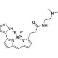
LysoTracker Red is a red-fluorescent lysosomal probe with excitation/emission maxima at 577/590 nm. It consists of a fluorophore linked to a weakly basic moiety, allowing it to freely cross cell membranes and selectively accumulate in acidic organelles such as lysosomes. LysoTracker Red is commonly used for visualizing lysosomal structures and studying lysosome-related biosynthesis and pathogenesis.
-
- Cat. No.
- A24762
- CAS No.
- 215247-93-1
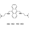
LysoTracker Blue DND-22 is a blue fluorescent dye designed for visualizing acidic cellular compartments. With an excitation/emission wavelength of 373/422 nm, this reagent effectively stains lysosomes in live cells. It is instrumental in research applications focused on neurodegenerative diseases and leukemia, facilitating the study of cellular processes and disease mechanisms associated with acidic organelles.
-
- Cat. No.
- A24866
- CAS No.
- 152584-38-8
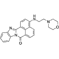
DND-189 is a low-pH fluorescent probe that exhibits high sensitivity to neutral and acidic pH environments. This reagent is valuable for the quantification of pH levels within acidic organelles, enabling researchers to study intracellular pH dynamics in various biological contexts. Its unique properties make DND-189 an essential tool for applications in cellular imaging and pH monitoring.
-
- Cat. No.
- A24918
- CAS No.
- 172971-76-5
Synonyms: IR-808
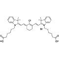
MHI-148 is a near-infrared heptamethine cyanine dye that selectively targets tumors, making it ideal for cancer detection and diagnosis. This compound demonstrates preferential accumulation in the lysosomes and mitochondria of tumor cells, allowing for enhanced visualization. Its unique properties facilitate research applications in tumor biology and imaging studies, enabling more effective monitoring and understanding of cancer progression.
-
- Cat. No.
- A24941
- CAS No.
- 220524-71-0
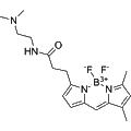
Green DND-26 is a lysosome-specific fluorescent dye that exhibits a maximum excitation/emission wavelength of 504/511 nm. Comprising a fluorescein moiety linked to weak bases, this compound effectively penetrates cell membranes and accumulates in lysosomal structures. It is ideal for studying lysosomal biosynthesis and related pathological processes, facilitating the observation of cellular dynamics associated with lysosomal function.
-
- Cat. No.
- A24974
- CAS No.
- 1064123-31-4
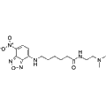
LysoTracker Yellow HCK 123 is a membrane-permeable fluorescent probe that selectively targets acidic cellular compartments. This compound is a weakly basic amine that accumulates in environments with low luminal pH, enabling researchers to visualize and study lysosomal dynamics and function. It is widely utilized in cellular imaging applications to investigate cellular processes such as autophagy, metabolism, and organelle dynamics.
-
- Cat. No.
- A39622
- CAS No.
- 2781892-92-8
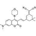
NCIC-VIS is a lysosome-targeted, viscosity-sensitive two-photon near-infrared fluorescent probe. Its rigid molecular structure minimizes torsional motion, enhancing the fluorescence quantum yield and two-photon absorption cross-section. NCIC-VIS facilitates real-time imaging of autophagy processes within cells, making it a valuable tool for researchers investigating cellular dynamics and lysosomal function.
-
- Cat. No.
- A43789
- CAS No.
- 2324152-35-2
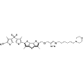
Lyso Flipper-TR 29 is a lysosomal flipper probe designed to selectively label lysosomes and late endosomes. This compound utilizes transient deprotonation to achieve membrane permeability in its neutral form. It serves as a valuable tool for researchers studying lysosomal dynamics, trafficking, and function in various cellular contexts. By employing Lyso Flipper-TR 29, investigators can gain insights into lysosomal biogenesis and turnover, making it suitable for applications in cell biology and related fields.
-
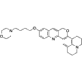
- Cat. No.
- A44866
- CAS No.
- 2089438-23-1
CQ-Lyso is a lysosomal pH probe that utilizes a chromenoquinoline structure to selectively target lysosomes in living cells. This fluorescent probe enables real-time measurement of lysosomal pH under single wavelength excitation, allowing researchers to visualize dynamic pH changes within lysosomes. CQ-Lyso is valuable for studying lysosomal function and pathology in various biological contexts.
-
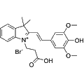
- Cat. No.
- A44909
- CAS No.
- 2962939-33-7
INSA is a pH-sensitive fluorescent probe that effectively distinguishes between the internal pH of mitochondria and lysosomes. This reagent emits red fluorescence in mitochondria and green fluorescence in lysosomes, allowing for simultaneous visualization of these organelles. INSA is particularly useful for studying cellular metabolism, organelle function, and intracellular pH dynamics in various biological research applications.
-
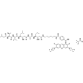
- Cat. No.
- A45282
- CAS No.
- 2410614-07-0
JF526–Pepstatin A TFA is a fluorescent dye designed for lysosomal staining in live cells. It exhibits an excitation maximum at 530 nm and an emission maximum at 549 nm, making it suitable for various fluorescence microscopy applications. This reagent facilitates the visualization of lysosomes, contributing to studies in cellular dynamics, drug delivery mechanisms, and autophagy processes.
-
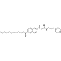
- Cat. No.
- B11166
- CAS No.
- 3091879-73-8
Lyso-laurdan is a fluorescent lipid probe that targets lysosomal membranes. It features a morpholine group that enables its accumulation in lysosomes through protonation, allowing it to adopt a cationic form in the acidic environment of these organelles. This localization facilitates the investigation of lysosomal membrane properties and dynamics, making it a valuable tool for researchers studying lysosomal function and related cellular processes.
-
- Cat. No.
- B11201
- CAS No.
- 2567068-39-5
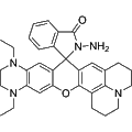
ONOO-LysopH is a Rhodamine-based near-infrared fluorescent dye specifically designed to target lysosomal pH and detect peroxynitrite (ONOO−). This probe exhibits a significant fluorescence enhancement at approximately 686 nm under acidic conditions, while showing a rapid NIR turn-on at around 678 nm in the presence of ONOO− at physiological pH. ONOO-LysopH is ideal for live cell imaging to monitor lysosomal pH fluctuations and to visualize endogenous or exogenous peroxynitrite levels, thereby providing valuable insights into cellular processes.
Selection Guide
Where is the signal?
LysoTracker Red DND-99 was used to visualize acidic vesicles in a glioblastoma-cell study. Acidotropic staining can include late endosomes as well as lysosomes, so use an appropriate localization strategy when the identity of the compartment matters. [1] [2]
Is a pH value required?
DND-189 is documented as LysoSensor Green DND-189 in the cited methods. One study converted its fluorescence to pH using sequential known-pH buffers and calibration under matched acquisition settings. An uncalibrated bright or dim image is not that measurement. [3]
Could loading or organelle changes affect the comparison?
For LysoTracker, total signal depends on factors including organelle size, number and contents. Separate compartment visualization from pH inference; do not apply a handling or fixation conclusion from a different dye variant without supporting evidence. [2]
Representative Reagent Comparison
Selected examples for this topic; independent of the current product page or filters.
Representative reagents and selection considerations
| Reagent / SKU | Documented use | What the signal supports | Method boundary |
|---|
| LysoTracker Red (LysoTracker Red DND-99)A24512 |
Acidic-vesicle visualization in U251N glioblastoma cells. | Localization of acidic compartments. | Not lysosome-exclusive and not a standalone pH meter; total staining can vary with organelle properties and dye accumulation. [1] [2] |
|---|
| DND-189A24866 |
Acidic-organelle pH-responsive fluorescence; published as LysoSensor Green DND-189. | pH inference when calibrated in the experimental system. | The cited cell study used known-pH buffers and matched imaging settings. Raw fluorescence is not an absolute pH value; do not transfer a DND-160 ratio method to DND-189. [3] |
|---|
A24512
- Documented use
- Acidic-vesicle visualization in U251N glioblastoma cells.
- What the signal supports
- Localization of acidic compartments.
- Method boundary
- Not lysosome-exclusive and not a standalone pH meter; total staining can vary with organelle properties and dye accumulation. [1] [2]
A24866
- Documented use
- Acidic-organelle pH-responsive fluorescence; published as LysoSensor Green DND-189.
- What the signal supports
- pH inference when calibrated in the experimental system.
- Method boundary
- The cited cell study used known-pH buffers and matched imaging settings. Raw fluorescence is not an absolute pH value; do not transfer a DND-160 ratio method to DND-189. [3]
The two examples represent different experimental choices, not interchangeable dyes. Confirm exact material and sample-processing conditions before adopting a method.
References
-
Maysinger D, Gran ER, Bertorelle F, Fakhouri H, Antoine R, Kaul ES, Samhadaneh DM, Stochaj U. Gold nanoclusters elicit homeostatic perturbations in glioblastoma cells and adaptive changes of lysosomes.
Theranostics. 2020. DOI: 10.7150/thno.37674. PMID: 32042327. PMCID: PMC6993243.
-
Barral DC, Staiano L, Guimas Almeida C, Cutler DF, Eden ER, Futter CE, Galione A, Marques ARA, Medina DL, Napolitano G, Settembre C, Vieira OV, Aerts JMFG, Atakpa-Adaji P, Bruno G, Capuozzo A, De Leonibus E, Di Malta C, Escrevente C, Esposito A, Grumati P, Hall MJ, Teodoro RO, Lopes SS, Luzio JP, Monfregola J, Montefusco S, Platt FM, Polishchuck R, De Risi M, Sambri I, Soldati C, Seabra MC. Current methods to analyze lysosome morphology, positioning, motility and function.
Traffic (Copenhagen, Denmark). 2022. DOI: 10.1111/tra.12839. PMID: 35343629. PMCID: PMC9323414.
-
Ghosh S, Dellibovi-Ragheb TA, Kerviel A, Pak E, Qiu Q, Fisher M, Takvorian PM, Bleck C, Hsu VW, Fehr AR, Perlman S, Achar SR, Straus MR, Whittaker GR, de Haan CAM, Kehrl J, Altan-Bonnet G, Altan-Bonnet N. β-Coronaviruses Use Lysosomes for Egress Instead of the Biosynthetic Secretory Pathway.
Cell. 2020. DOI: 10.1016/j.cell.2020.10.039. PMID: 33157038. PMCID: PMC7590812.
