Search by product name, CAS, target or catalog number. Press Down Arrow to reach product suggestions, Tab to move between links, and Escape to close suggestions.
USD – US Dollar✓Current currency
Account
Cell Viability & Live/Dead Stains
Choose a reagent by the cellular property you need to assess: membrane integrity, intracellular enzyme activity or metabolic reducing activity. These readouts answer different questions and should not be treated as interchangeable measures of survival. [1][2][3] Compare the selected reagents below, then match the assay to your sample, treatment and detection method. Use complementary measurements when a single signal cannot resolve the biological question.
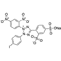
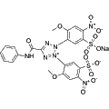
WST-3 is a water-soluble tetrazolium dye that serves as a fluorescent probe for cellular activity. Upon reduction by NADH in the presence of an electron mediator, WST-3 is converted to WST-3 formazan, which exhibits a maximum absorption at 433 nm. This dye is widely utilized in cell viability assays and metabolic activity studies, making it a valuable tool for various biological and pharmaceutical research applications.
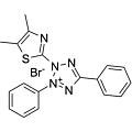
Synonyms: MTT; Thiazolyl Blue Tetrazolium bromide; Methylthiazolyldiphenyl-tetrazolium bromide
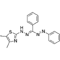
Thiazolyl Blue (MTT) is a colorimetric agent widely used to measure cell proliferation. Thiazolyl Blue (MTT) is reduced from yellow color to purple formazan in living cells.
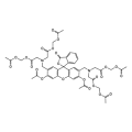
Calcein AM is a cell-permeable, non-fluorescent dye that readily enters live cells. Once inside, it is hydrolyzed by intracellular esterases to generate Calcein, a polar and highly negatively charged molecule that is retained within the cytoplasm. Calcein emits strong green fluorescence (Ex/Em: 494/515 nm) and is commonly used in combination with Propidium Iodide for assessing cell viability and cytotoxicity.
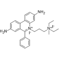
Propidium Iodide (PI) is a nuclear staining agent that stains DNA. Propidium Iodide is an analogue of ethidine bromide that emits red fluorescence upon embedding in double-stranded DNA. Propidium Iodide cannot pass through living cell membranes, but it can pass through damaged cell membranes to stain the nucleus. Propidium Iodide has a fluorescence wavelength of 493/617 nm and a wavelength of 536/635 nm after Mosaic with DNA. Propidium Iodide is commonly used in the detection of apoptosis (apoptosis) or necrosis (necrosis), and is often used in flow cytometry analysis.
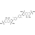
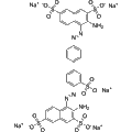
Synonyms: Direct Blue 14; Diphenyl Blue; Diamine Blue
Diphenyl Blue, also known as Trypan Blue, is a vital reference dye primarily used to assess cell membrane integrity and viability. It selectively stains dead or damaged cells blue, allowing for the identification of non-viable cells in various tissue and cell culture applications. Viable cells with intact membranes exclude the dye, while macrophages can phagocytize Diphenyl Blue, making it an effective living stain for studying these immune cells. This dye is essential for research involving cell viability assays and tissue analyses.
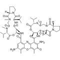
7-Aminoactinomycin D (7-AAD) is a cell-impermeant fluorescent DNA stain that primarily targets and binds to GC-rich regions of DNA. This compound is often utilized in flow cytometry and fluorescence microscopy to assess cell viability and apoptosis. Additionally, 7-AAD exhibits antibacterial properties, expanding its utility in various biological research applications.
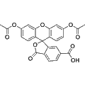
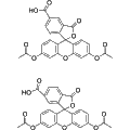
6-CFDA is a fluorescent dye commonly used in biological research for cell tracking and viability assays. It undergoes free diffusion into cells, where it is hydrolyzed by intracellular non-specific lipases to produce carboxyfluorescein, a fluorescent compound that carries an additional negative charge. This enhanced charge improves retention within cells compared to traditional fluorescein dyes, facilitating more accurate analysis of cellular processes and functions.
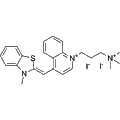
NIR-Red Dead Cell-1 Dye is a DNA-binding fluorochrome designed for the detection of necrotic and late apoptotic cells by intercalating into double-stranded DNA. It emits strong green fluorescence (Ex/Em=515 nm/531 nm) when bound to damaged cell membranes, making it an effective tool for distinguishing live cells from dead cells in fluorescence microscopy and flow cytometry applications. Additionally, NIR-Red Dead Cell-1 Dye can be conjugated to Feraheme nanoparticles, facilitating its use in drug delivery studies by providing fluorescent labeling.
Trypan red is a vital stain primarily used for the assessment of cell viability. It selectively enters dead cells, allowing researchers to differentiate live and non-viable cells in various biological samples. This reagent is widely utilized in cell culture, toxicity assays, and immunological studies to evaluate cell health and proliferation.
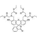
Calcein (tetraethyl ester) is a fluorescent dye that permeates cellular membranes, enabling its use in live cell imaging. This compound is widely utilized for assessing cell viability and for short-term labeling applications in biological research. Its strong fluorescence allows for effective visualization of cellular processes in various experimental contexts.
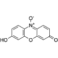
Resazurin is a water-soluble, membrane-permeable fluorescent dye that serves as a redox indicator. It is non-toxic and stable, transitioning from a blue non-fluorescent form to a pink and highly fluorescent form, Resorufin, upon reduction in living cells. Due to its sensitivity to changes in cellular metabolism, Resazurin is widely used for assessing cell viability, cytotoxicity, proliferation, migration, and invasion across various biological systems, including human, plant, animal, bacterial, and fungal cells.
NIR-Red Dead Cell-3 Dye is a specialized dye with a primary mechanism for labeling and visualizing dead cells in biological samples. This reagent is essential for various applications, including monitoring cell viability, analyzing cellular structures, and distinguishing between live and dead cell populations. Its versatility makes it suitable for use in both basic research and clinical diagnostics, facilitating insights into cell behavior and tissue pathology. Additionally, it can be applied in emerging technologies, such as functional textiles and dye-sensitized solar cells.
5(6)-CFDA is a fluorescent dye that acts through passive diffusion into cells, where it is hydrolyzed by intracellular non-specific lipases into carboxyl fluorescein (CF). The carboxylate group of CF imparts a negative charge, enhancing its retention within cells compared to traditional fluorescein dyes. This property makes 5(6)-CFDA particularly useful for various biological applications such as cell tracking, apoptosis detection, and cellular viability assays in live cell imaging studies.
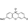
Resazurin sodium is a non-toxic, stable redox indicator that targets bacterial activity through its membrane-permeable properties. It serves as a vital tool for assessing cell viability, proliferation, and toxicity in various cell types, including human, plant, animal, bacterial, and fungal cells. Upon reduction, Resazurin sodium transforms into Resorufin, a highly fluorescent compound, enabling sensitive detection and quantification of cellular processes under fluorescent microscopy or spectrophotometry.
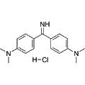
Auramine O is a fluorescent dye primarily used for the detection of amyloid fibrils. It exhibits strong yellow fluorescence, making it valuable for visualizing cellular structures and assessing algal cell viability. Additionally, Auramine O is employed in staining acid-fast bacteria, thereby aiding in microbiological studies and cancer research involving lung malignancies.
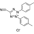
5-Cyano-2,3-di-(p-tolyl)tetrazolium chloride is a redox-sensitive fluorescent dye primarily used as a metabolic activity indicator in microorganisms. This compound emits red fluorescence with a maximum emission at 602 nm, enabling effective visualization and quantification of cellular metabolism. It serves as a valuable tool in microbiological studies, particularly for assessing viability and metabolic function in various microbial species.
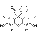
Eosin Y (Acid Red 87 free base) is a soluble acid dye known for its role as a photoredox catalyst. This dye exhibits strong absorbance in the visible light spectrum, facilitating various organic synthesis reactions. Eosin Y is commonly utilized in photochemical applications, enabling the generation of reactive intermediates for diverse chemical transformations.
SYTO9 is a green fluorescent nucleotide dye that selectively binds to nucleic acids, with excitation at 485 nm and emission at 498 nm for DNA and 501 nm for RNA. This dye exhibits the ability to penetrate intact cell membranes of viable bacteria, with increased permeability observed in compromised membranes. SYTO9 is commonly utilized in fluorescence microscopy, flow cytometry, and fluorometric assays in microbiology, enabling applications in bacterial viability assessment, cell imaging, and quantification of microbial populations.
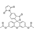
Fluorescein diacetate 5-maleimide is a fluorescent dye that serves as a versatile labeling agent for biomolecules. It exhibits strong fluorescence upon hydrolysis, facilitating the detection of cellular components and the study of cellular viability. This compound is particularly useful in applications involving flow cytometry, live-cell imaging, and tracking molecular interactions in various biological research contexts.
XTT Sodium is a tetrazolium dye primarily utilized for assessing cell viability based on redox potential. Actively respiring cells reduce XTT, resulting in the formation of a water-soluble orange-colored formazan product. This reagent is commonly employed in cytotoxicity assays and proliferation studies to quantify cellular metabolic activity.
MTT Formazan, a purple water-insoluble compound, is generated through the reduction of yellow soluble MTT by succinate dehydrogenase in the mitochondria of viable cells. This product serves as a reliable indicator of cellular metabolic activity, making it essential for evaluating cell viability and proliferation. The absorbance of MTT Formazan can be quantitated via spectrophotometry, providing insight into cellular status. Additionally, it is applicable in bacterial MTT assays and enhances imaging in photoacoustic microscopy, particularly in three-dimensional cell culture and tissue engineering studies.
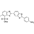
Primulin is a fluorescent dye that exhibits strong binding affinity for albumin, making it a valuable tool in various analytical and biological studies. This compound effectively labels plant cell walls and distinguishes between live and dead spermatozoa through distinct fluorescence patterns. In neurobiology, Primulin serves as a retrograde axonal tracer, while its derivatives demonstrate inhibitory activity against HCV NS3 and interference with dengue virus NS3-mediated ATP hydrolysis. Additionally, Primulin disrupts HCV replicase assembly, highlighting its potential in virology research applications.
Calcein-AM becomes fluorescent after esterase conversion, whereas propidium iodide uptake can reveal loss of membrane exclusion. Human trabecular-meshwork experiments used both reagents with viable and damaged-tissue controls. That example supports complementary readouts, not a universal threshold for every sample. [1][2]
Could the treatment change the reporter?
Calcein retention depends on cellular conditions as well as conversion. Resazurin reports reducing activity, which can change without demonstrating cell death. Include appropriate assay controls and account for non-cellular reduction when interpreting metabolic assays. [1][3]
What conclusion is needed?
Distinguish an esterase/retention signal, a membrane-integrity observation and a metabolic comparison in your analysis. Do not rename all three as a direct count of living cells. The selected examples do not establish an apoptosis mechanism or a proliferation rate. [1][2][3]
Representative Reagent Comparison
Selected examples for this topic; independent of the current product page or filters.
Representative reagents and selection considerations
Reduction to resorufin; fluorescence or absorbance readout.
Metabolic reducing activity rather than direct death staining.
The paper supports resazurin assay principles, not SKU-specific performance. Non-cellular reduction and nonlinear response can bias interpretation. [3]
Reduction to resorufin; fluorescence or absorbance readout.
Useful distinction
Metabolic reducing activity rather than direct death staining.
Experimental boundary
The paper supports resazurin assay principles, not SKU-specific performance. Non-cellular reduction and nonlinear response can bias interpretation. [3]
Use the exact reagent form and controls appropriate to your model. The comparison is not a survival ranking or an Adooq experimental validation.