
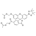
- Cat. No.
- A14086
PF6-AM
Synonyms: Peroxyfluor-6 AM
PF6-AM is a new selective fluorescent indicator for hydrogen peroxide (H(2)O(2)), a tool to help discover new redox chemistry in biological systems.
Out of stock
All 3 sizesSearch by product name, CAS, target or catalog number. Press Down Arrow to reach product suggestions, Tab to move between links, and Escape to close suggestions.
Select an oxidative-stress probe by the reaction and cellular compartment you need to investigate, not by the label “ROS” alone. DCF-based fluorescence, mitochondria-associated probe oxidation and membrane-associated oxidation provide different kinds of information. [1] [2] The selected reagents illustrate these choices. Interpret signal changes alongside method-specific controls; fluorescence intensity alone may not identify a particular oxidant or quantify oxidative damage. [1] [3]
Compare selected reagents
Synonyms: Peroxyfluor-6 AM
PF6-AM is a new selective fluorescent indicator for hydrogen peroxide (H(2)O(2)), a tool to help discover new redox chemistry in biological systems.
Out of stock
All 3 sizes
BODIPY 581/591 C11 is a boron-dipyrromethene (BODIPY) derivative with excellent photostability and minimal fluorescence artifacts. It is widely used to study lipid peroxidation and antioxidant activity in living cells, and to detect ferroptosis through its reaction with hydroxyl radicals. The dye exhibits a fluorescence shift upon oxidation: the reduced form emits at 591 nm (Ex: 581 nm), while the oxidized form emits at 510 nm (Ex: 500 nm).
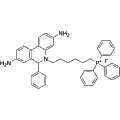
MitoSOX Red is a live cell fluorescent probe that specifically targets mitochondria and is cell membrane permeable. MitoSOX Red enters mitochondria and is oxidized by superoxide but not by other ROS or RNS generating systems. The oxidized MitoSOX Red then binds to nucleic acids in mitochondria/nucleus, producing strong red fluorescence. MitoSOX Red can be used as a fluorescent indicator to specifically detect superoxide. In addition, superoxide dismutase (SOD) can prevent the oxidation of MitoSOX Red. Excitation/emission wavelength: 510/580 nm.
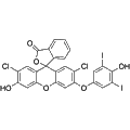
HKOH-1 is a sensitive green fluorescent probe designed for the selective detection of hydroxyl radicals (·OH) in living cells, utilizing a maximum excitation wavelength of 500 nm and an emission wavelength of 520 nm. This reagent enables researchers to monitor oxidative stress and assess cellular responses to reactive oxygen species. Its application is critical in studies related to redox biology, neurodegenerative diseases, and cancer research.
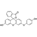
Synonyms: HPF; 3'-p-(Hydroxyphenyl) fluorescein
Hydroxyphenyl Fluorescein (HPF) functions as a stable fluorescent probe specifically targeting reactive oxygen species (ROS). This compound exhibits enhanced specificity and stability compared to traditional probes such as H2DCFDA. It generates strong green fluorescence upon reaction with intracellular peroxynitroso, making it suitable for studying oxidative stress. Hydroxyphenyl Fluorescein can be utilized in various applications, including fluorescence microscopy, high-throughput imaging, luciferase microplate assays, and flow cytometry. Excitation and emission wavelengths are 490 nm and 515 nm, respectively.
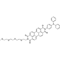
LPd peroxida probe is a fluorescent marker specifically designed to detect lipid peroxidation, a key process in ferroptosis. This probe effectively converts lipid hydroperoxides into lipid alcohols, facilitating the imaging of lipid hydroperoxides in live cells. It is an invaluable tool for researchers exploring the implications of lipid peroxidation in various cellular pathophysiologies.
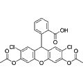
Synonyms: DCFH-DA; 2',7'-Dichlorodihydrofluorescein diacetate
H2DCFDA is a cell-permeable probe designed for the detection of intracellular reactive oxygen species (ROS). This fluorescent indicator exhibits excitation and emission maxima at 488 nm and 525 nm, respectively. H2DCFDA is widely utilized in research applications involving oxidative stress and cellular signaling, providing insights into ROS-related biological processes.
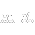
CM-H2DCFDA is a cell-permeable fluorescent probe designed for the detection of reactive oxygen species (ROS) within cells. Upon oxidation by ROS, this compound releases a highly fluorescent product, facilitating the measurement of cellular oxidant levels with an excitation/emission maximum of 495/530 nm. Suitable for various applications in oxidative stress research, CM-H2DCFDA provides sensitive and reliable quantification of ROS in live-cell imaging studies. Note that this reagent is light-sensitive and should be handled accordingly.
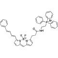
MitoPerOx is a fluorescent probe designed to detect lipid peroxidation specifically in mitochondria, utilizing BODIPY581/591 fluorophores. With its triphenylphosphine cation (TPP+), MitoPerOx selectively accumulates in mitochondria based on membrane potential, allowing for sensitive measurement of lipid peroxides within the inner mitochondrial membrane. The probe exhibits a ratiometric emission shift from 590 nm to 520 nm upon oxidation, facilitating accurate detection at an excitation wavelength of 488 nm. MitoPerOx is valuable for investigating oxidative stress-associated diseases, including aging, neurodegenerative disorders, and mitochondrial dysfunction.
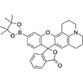
Synonyms: PO1
Peroxy Orange 1 is a hydrogen peroxide (H2O2) specific probe designed for the detection of reactive oxygen species. It binds selectively to highly reactive oxygen species, enabling the visualization of H2O2 in live cells through green fluorescence. This compound is valuable for studying oxidative stress, cellular signaling, and related biological processes in various research applications.
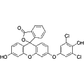
HKOCl-3 is a selective fluorescent probe designed for the detection of hypochlorous acid. This reagent exhibits high sensitivity with an excitation wavelength of 490 nm and an emission wavelength of 527 nm. HKOCl-3 is suitable for various biological research applications involving oxidative stress, inflammation studies, and the monitoring of hypochlorous acid levels in biological samples.
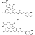
HKPerox-2 is a highly sensitive fluorescent probe specifically designed for the detection of hydrogen peroxide (H2O2) in live cells. It exhibits peak excitation and emission wavelengths at 520 nm and 543 nm, respectively, making it suitable for fluorescence microscopy applications. This reagent can be utilized to study oxidative stress and cellular signaling involving reactive oxygen species, enabling insights into various biological processes and disease mechanisms.
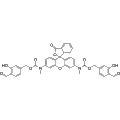
HKPerox-1 is a highly sensitive green fluorescent probe designed for the specific detection of hydrogen peroxide (H2O2) in living cells. It exhibits a maximum excitation wavelength of 520 nm and an emission wavelength of 543 nm, enabling effective visualization of oxidative stress and reactive oxygen species. This probe is ideal for research applications involving cellular metabolism, oxidative stress studies, and the assessment of redox biology in various experimental contexts.
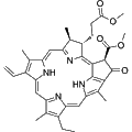
Methyl pheophorbide a is a chlorophyll-a derivative that functions as a potent photosensitizer. Upon exposure to specific wavelengths of light, it generates singlet oxygen and reactive oxygen species (ROS), inducing cytotoxic effects and promoting cell death. This compound is commonly utilized in photodynamic therapy research and studies focused on oxidative stress and cellular response mechanisms.
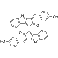
Scytonemin is a hydrophobic alkaloid pigment derived from the outer sheath of cyanobacteria, primarily functioning as a natural ultraviolet sunscreen. This compound exhibits significant protective properties against short-wave solar ultraviolet (UV) radiation, thereby reducing the generation of reactive oxygen species (ROS) and mitigating DNA damage. Additionally, Scytonemin displays anti-inflammatory and anti-proliferative effects, with a concentration-dependent inhibition of polo-like kinase 1 (PLK1)-mediated cdc25C phosphorylation (IC50 = 2.0 μM). This makes it a valuable reagent for research in cancer biology, acute inflammation, and the development of sunscreen cosmetics.
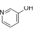
3-Hydroxypyridine is a natural photosensitizer that facilitates oxidative stress and influences cell proliferation and apoptosis in skin cells upon UVA/UVB irradiation. This compound, which can be derived from bamboo grass, plays a significant role in skin biology and phototoxicity research. Its ability to mediate cellular responses under light exposure makes it a valuable tool for studies related to skin damage and photoaging.
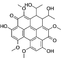
Elsinochrome C is a photosensitizer that activates upon light exposure to generate reactive oxygen species (ROS), including singlet oxygen (1O2). This mechanism induces apoptosis and disrupts pathogen structures, making it a valuable tool in cancer research and the study of HIV-associated infections. Additionally, Elsinochrome C shows potential in investigating refractory skin diseases.
Out of stock
All 3 sizes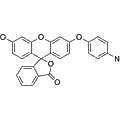
APF is a cell-permeable fluorescence probe designed for the detection of hydroxyl radicals within reactive oxygen species (ROS). This compound is inherently nonfluorescent and produces a distinct bright green fluorescence upon interaction with hydroxyl radicals. APF is suitable for measuring intracellular ROS levels, with optimal excitation and emission at 490 nm and 525 nm, respectively, making it a valuable tool for various biological and biochemical research applications.
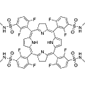
Synonyms: LUZ11; F2BMet
Redaporfin is a potent photosensitizer that generates reactive oxygen species upon light activation. This compound exhibits direct antineoplastic effects and enhances immune-mediated destruction of malignant lesions, making it valuable for cancer treatment research. Its application in photodynamic therapy allows for targeted approaches in oncology, promising improved outcomes in the treatment of various tumors.
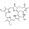
Chlorin e6 trimethyl ester is a potent photosensitizer derived from methyl pheophorbide-a, primarily utilized in photodynamic therapy (PDT). This compound demonstrates significant biological activity by generating reactive oxygen species upon light activation, which induces cell death in malignant tissues. Research applications include cancer treatment, where targeted phototherapy can enhance therapeutic effectiveness while minimizing damage to surrounding healthy cells.
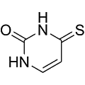
4-Thiouracil is a thionucleobase recognized for its cytostatic properties and function as a UVA photosensitizer. This compound can be utilized as a biological photoprobe to investigate RNA structures and nucleic acid interactions. Additionally, 4-Thiouracil generates reactive oxygen species, such as O2, upon UVA exposure, making it valuable for research in photocrosslinking, photodamage, and photodynamic therapy applications.
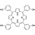
Synonyms: Tetra(3-hydroxyphenyl)porphyrin; 3-THPP
m-THPP (Tetra(3-hydroxyphenyl)porphyrin) functions as a photosensitizer, exhibiting the ability to induce reactive oxygen species (ROS) production. It demonstrates notable antitumor and antibacterial activities, making it a valuable reagent for photodynamic therapy (PDT) research. Its applications span various studies aimed at exploring therapeutic strategies against cancer and microbial infections.
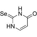
2-Selenouracil is a specialized photosensitizer that targets cellular components in photodynamic therapy applications. It exhibits significant biological activity, effectively inducing cytotoxic effects upon exposure to light. This compound is primarily utilized in cancer research and treatment protocols, particularly for its ability to generate reactive oxygen species (ROS) that can selectively destroy cancerous cells.
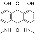
Disperse Polyester Dark Blue is a redox-active probe designed for the identification of reactive oxygen species (ROS) in biological samples. It operates by accepting electrons from ROS, undergoing oxidation, and subsequently generating a measurable chemiluminescent signal. This signal intensity directly correlates with ROS levels, making it a valuable tool for researchers investigating oxidative stress and related pathways in cells and tissues.
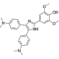
Photosensitizer-1 is a potent chemical agent that acts as a photosensitizer, facilitating photodynamic therapy applications. By generating reactive oxygen species upon light activation, it effectively targets and induces cell death in various malignancies. This compound is valuable for research in cancer therapy and studying the mechanisms of photodynamic treatments.
H2DCFDA supports oxidation-dependent DCF measurements; it is not a direct hydrogen-peroxide concentration assay. MitoSOX is used in mitochondrial oxidation studies, but different oxidation products contribute overlapping fluorescence. [1]
A red MitoSOX signal alone does not establish superoxide production. Product separation and identification are needed for that attribution. DCF chemistry can also be affected by heme proteins, metals and signal amplification reactions; use a method that can address these confounders. [1]
C11-BODIPY has documented use as an oxidation-sensitive membrane probe. Its response should not be equated with the absolute amount of endogenous lipid oxidation: a comparison with mass spectrometry found overreporting and perturbation of oxidation in the tested system. Treat it as an indicator within a validated assay. [2] [3]
Selected examples for this topic; independent of the current product page or filters.
| Reagent / SKU | Documented readout | Selection use | Key interpretation limit |
|---|---|---|---|
| H2DCFDAA24745 | Oxidation-dependent DCF fluorescence after cellular processing. | Broad redox-related changes. | Does not react directly with H2O2 to produce DCF; signal is not a specific oxidant concentration. [1] |
| MitoSOX RedA24559 | Mitochondria-associated oxidation products. | Investigating mitochondrial oxidant-related processes. | Total red fluorescence mixes products; identify the diagnostic hydroxylated product before attributing the signal to superoxide. [1] |
| BODIPY 581/591 C11A24539 | Oxidation-sensitive C11-BODIPY response in membrane systems and cells. | Membrane-associated oxidation / lipid-peroxidation studies. | Not an absolute lipid-oxidation measurement. The dye overreported and inhibited oxidative damage in a model-membrane comparison. [2] [3] |
These probes have useful, conditional research roles. A signal change is not automatically a specific ROS concentration or proof of a particular cell-death pathway.
Free radical biology & medicine. 2012. DOI: 10.1016/j.freeradbiomed.2011.09.030. PMID: 22027063. PMCID: PMC3911769.
Free radical biology & medicine. 2002. DOI: 10.1016/s0891-5849(02)00848-1. PMID: 12160930.
Free radical biology & medicine. 2007. DOI: 10.1016/j.freeradbiomed.2007.01.038. PMID: 17395012.