
- Cat. No.
- A15135
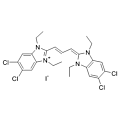
JC-1
Synonyms: CBIC2
JC-1 is a dual-emission potential-sensitive probe that can be used to measure mitochondrial membrane potential.
In stock
View Details
Search by product name, CAS, target or catalog number. Press Down Arrow to reach product suggestions, Tab to move between links, and Escape to close suggestions.
Choose a mitochondrial reagent according to the readout you need: mitochondrial localization or a change in membrane potential. JC-1 aggregation and TMRE accumulation provide potential-related signals, while MitoTracker Green FM offers a localization example. [1] [2] [3] These signals require method-specific interpretation. A change in fluorescence alone is not a direct count of mitochondria; consider dye behavior, sample handling and controls before comparing samples. [1] [2] [3]
Compare selected reagents
Synonyms: CBIC2
JC-1 is a dual-emission potential-sensitive probe that can be used to measure mitochondrial membrane potential.
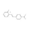
2-Di-1-ASP (Compound 18a) is a mono-stryryl dye, and widely used as mitochondrial stain and groove-binding fluorescent probes for double-stranded DNA. 2-Di-1-ASP is selective for G-quadruplex (G4) and double-stranded DNA.
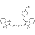
MitoTracker Deep Red FM is a fluorescent dye that selectively accumulates in the mitochondrial matrix and covalently binds to mitochondrial proteins by reacting with free thiol groups of cysteine residues. This allows for stable mitochondrial labeling independent of membrane potential. The dye has excitation/emission maxima at 644/665 nm. Store protected from light.
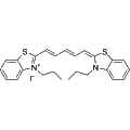
DiSC3(5) is a fluorescent probe commonly used to assess mitochondrial membrane potential, with excitation/emission maxima at 622/670 nm. It can inhibit mitochondrial NAD-linked respiratory activity, with an IC₅₀ of 8 μM. In the presence of the Na⁺/K⁺-ATPase inhibitor ouabain, DiSC3(5) induces membrane hyperpolarization in Ehrlich ascites tumor cells.
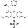
Synonyms: T668
Rhodamine dyes are membrane-permeable, cationic fluorescent probes that selectively accumulate in mitochondria by recognizing mitochondrial membrane potential, producing bright fluorescence. At appropriate concentrations, they exhibit low cytotoxicity and are widely used for mitochondrial staining in animal cells, plant cells, and microorganisms.
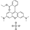
Synonyms: Tetramethylrhodamine ethyl ester perchlorate
Rhodamine dyes are membrane-permeable, cationic fluorescent probes that selectively accumulate in mitochondria by sensing mitochondrial membrane potential, resulting in bright fluorescence. Due to their low cytotoxicity at appropriate concentrations, they are widely used for mitochondrial labeling in animal cells, plant cells, and microorganisms.
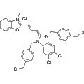
MitoTracker Green FM is a green fluorescent dye that selectively targets the mitochondrial matrix. It covalently binds to mitochondrial proteins through reaction with free mercaptan groups on cysteine residues, facilitating the visualization of mitochondrial structures independently of membrane potential. This dye is widely utilized in cellular and molecular biology research to study mitochondrial dynamics, function, and viability. Excitation and emission wavelengths are 490 nm and 523 nm, respectively.
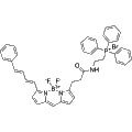
MitoPerOx is a fluorescent probe designed to detect lipid peroxidation specifically in mitochondria, utilizing BODIPY581/591 fluorophores. With its triphenylphosphine cation (TPP+), MitoPerOx selectively accumulates in mitochondria based on membrane potential, allowing for sensitive measurement of lipid peroxides within the inner mitochondrial membrane. The probe exhibits a ratiometric emission shift from 590 nm to 520 nm upon oxidation, facilitating accurate detection at an excitation wavelength of 488 nm. MitoPerOx is valuable for investigating oxidative stress-associated diseases, including aging, neurodegenerative disorders, and mitochondrial dysfunction.
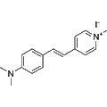
4-Di-1-ASP is a styryl dye that targets glioma cells for fluorescent imaging in live brain tissue. This reagent enables detailed analysis of cellular structures, viability, proliferation, and endocytic processes, including cytokinesis and phagocytosis. Additionally, 4-Di-1-ASP facilitates observations of mitochondrial architecture in living cells, exhibiting green fluorescence upon excitation (λex /λem = 475/606 nm).
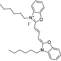
Synonyms: DiOC6(3) iodide
3,3'-Dihexyloxacarbocyanine iodide is a carbocyanine fluorescent dye primarily used for the assessment of mitochondrial membrane potential. This dye exhibits significant fluorescence characteristics, making it an effective tool for studying mitochondrial dynamics and function in various cellular processes. It is widely utilized in research applications involving cell viability, apoptosis studies, and mitochondrial health assessments.
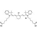
Synonyms: IR-808
MHI-148 is a near-infrared heptamethine cyanine dye that selectively targets tumors, making it ideal for cancer detection and diagnosis. This compound demonstrates preferential accumulation in the lysosomes and mitochondria of tumor cells, allowing for enhanced visualization. Its unique properties facilitate research applications in tumor biology and imaging studies, enabling more effective monitoring and understanding of cancer progression.
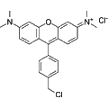
MitoTracker Orange CMTMRos is a fluorescent dye that selectively labels mitochondria in live cells by exploiting the mitochondrial membrane potential (Ex/Em: 551/576 nm). This compound is essential for studying mitochondrial dynamics, morphology, and function. It is widely used in research applications, including cellular metabolism, apoptosis, and drug-response assays in various cell types.
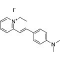
DASPEI is a cationic styrenyl dye that targets mitochondria, functioning as a fluorescent probe with a large Stokes shift. Exhibiting excitation and emission wavelengths of 550 nm and 573 nm respectively, DASPEI demonstrates excellent photostability and labeling capability. This reagent effectively stains mitochondria in living cells and can also label presynaptic nerve endings, providing insights into cellular and synaptic activity. Its applications are valuable in various fields including neurobiology and cellular imaging research.
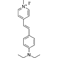
4-Di-2-ASP is a styryl pyridinium fluorescent dye primarily used as a mitochondrial marker. This compound demonstrates reliable and specific labeling of neuroepithelial bodies (NEBs) in pulmonary tissues, facilitating the investigation of mitochondrial dynamics and cellular respiration. Its application is critical in studies aimed at understanding respiratory physiology and neuroepithelial signaling.
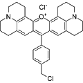
MitoMark Red I is a fluorescent dye specifically designed to target mitochondria in viable cells. This red fluorescent marker exhibits a peak excitation wavelength of 578 nm and an emission at 599 nm, enabling precise visualization of mitochondrial activity and dynamics. It is widely utilized in cellular and molecular biology research to study mitochondrial function, cell viability, and related physiological processes.
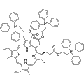
TPP-Ce6 is a mitochondria-targeting photosensitizer formed by conjugating chlorophyll e6 (Ce6) with triphenylphosphine (TPP) through an ester bond. Upon light irradiation, TPP-Ce6 generates reactive oxygen species (ROS), predominantly singlet oxygen, which can induce oxidative stress, resulting in mitochondrial dysfunction and apoptosis. This compound is useful for investigating mitochondrial roles in apoptosis and energy metabolism, as well as for applications in cancer therapy.
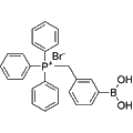
MitoB bromide is a mitochondria-targeted ratiometric probe designed to detect mitochondrial hydrogen peroxide levels. This compound acts as an exomarker for reactive oxygen species (ROS), allowing for the measurement of hydrogen peroxide in living organisms. It is valuable in research applications focused on oxidative stress and mitochondrial function, facilitating studies on cellular signaling and disease mechanisms involving ROS.
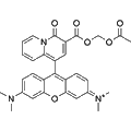
KMG-301AM is an acetoxymethyl ester of KMG-301 designed as a Mg2+ fluorescent probe targeting mitochondria. Upon accumulation within mitochondria, KMG-301AM is hydrolyzed to the active form, KMG-301, which is selective for Mg2+ detection in intact cells. This compound remains retained in the mitochondria due to the impermeable nature of the mitochondrial membrane, allowing it to reliably indicate variations in mitochondrial Mg2+ concentration and facilitate studies of Mg2+ transport across the mitochondrial membrane in cellular models.
Out of stock
All 3 sizes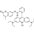
Mito Red is a vital dye that selectively accumulates in mitochondria, serving as a mitochondrial stain for evaluating mitochondrial function and status. Its fluorescence intensity is directly correlated with the mitochondrial membrane potential; an increase in membrane potential results in a corresponding increase in fluorescence signal. Mito Red is an essential tool for researchers studying mitochondrial activity, dysfunction, and related cellular processes.
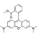
TMRM (Tetramethylrhodamine Methyl Ester) is a membrane-permeable cationic fluorescent dye that selectively targets mitochondrial membrane potential. Its ability to accumulate in mitochondria results in strong fluorescence, enabling the visualization of mitochondrial dynamics. TMRM is widely utilized in cellular research, including studies of mitochondrial function, cell viability, and apoptosis in various biological systems, including animal, plant, and microbial cells.
Out of stock
All 3 sizes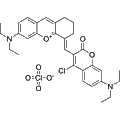
MI-BP-CC is a mitochondrial-targeting near-infrared fluorescent probe that localizes specifically within the mitochondria of live cells. It operates by visualizing mitochondrial viscosity through its fluorescent emission at 722 nm. This compound is useful for studies investigating mitochondrial function and dynamics in cellular environments.
Out of stock
All 3 sizes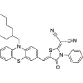
MIT-PZR is a mitochondria-targeted fluorescent probe designed for effective imaging applications. With low cytotoxicity, it is suitable for use in live-cell experiments and in vivo imaging studies. This probe exhibits excitation and emission maxima at 485 nm and 705 nm, respectively, enabling the visualization of mitochondrial activity and dynamics in real-time.
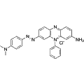
Janus Green is a cationic fluorescent dye primarily used for cellular and tissue staining. It exhibits strong affinity for mitochondria, allowing for visualization of these organelles in live and fixed cells. This dye is widely utilized in biological research to study mitochondrial morphology, function, and dynamics in various cell types. Janus Green is particularly valuable in experiments involving cell viability and apoptosis.
Out of stock
All 3 sizes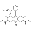
Synonyms: Basic Red 1
Rhodamine 6G is a cationic fluorescent dye that targets mitochondrial membrane potentials. It specifically localizes to mitochondria, emitting bright fluorescence, making it an effective tool for visualizing mitochondrial dynamics in various biological systems. With low toxicity at certain concentrations, Rhodamine 6G is widely employed in research applications involving animal cells, plant cells, and microorganisms to study mitochondrial function and health.
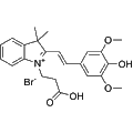
INSA is a pH-sensitive fluorescent probe that effectively distinguishes between the internal pH of mitochondria and lysosomes. This reagent emits red fluorescence in mitochondria and green fluorescence in lysosomes, allowing for simultaneous visualization of these organelles. INSA is particularly useful for studying cellular metabolism, organelle function, and intracellular pH dynamics in various biological research applications.
JC-1 changes between green monomer and red aggregate signals. TMRE responds through accumulation, but the cited isolated-mitochondria study also found quenching, binding and effects on respiration. Do not assume that brighter always means a higher potential. [1] [2]
Use the cited experimental context to plan controls and interpretation, rather than transferring a result between dyes or cell types. Green FM results depended on cell condition and removal of unbound dye before confocal imaging. [3]
The Green FM example here concerns living cells and tissues. It does not establish a general fixed-cell workflow. Check the intended processing sequence separately; no blanket claim about aldehyde retention is made here. [3]
Selected examples for this topic; independent of the current product page or filters.
| Reagent / SKU | Literature-supported use | Readout / interpretation | Experimental boundary |
|---|---|---|---|
| JC-1A15135 | Membrane-potential-related measurements. | Green monomer and red aggregate signals; aggregation also depends on dye concentration. | The cited characterization used buffers and isolated cardiac mitochondria. Interpret both signals with appropriate controls, not as a mitochondrial count. [1] |
| TMREA24538 | Membrane-potential assessment in isolated rat-heart mitochondria. | Potential-sensitive accumulation; fluorescence can be quenched after accumulation. | Membrane binding and respiratory effects affected interpretation. Use a TMRE-specific method; TMRM conditions are not interchangeable. [2] |
| MitoTracker Green FMA24612 | Mitochondrial localization in living cells and tissues. | Green fluorescence; relative insensitivity to potential was conditional in the cited study. | Cell damage and unbound dye affected readings. No general fixation-retention claim or direct mitochondrial-count calibration is established here. [3] |
These examples are not a ranking or a statement of interchangeability. Match the reagent, sample preparation and controls to the intended readout.
Biochemistry. 1991. DOI: 10.1021/bi00232a015. PMID: 2021638.
Biophys J. 1999. DOI: 10.1016/s0006-3495(99)77214-0. PMID: 9876159. PMCID: PMC1302536.
Cytometry A. 2004. DOI: 10.1002/cyto.a.20033. PMID: 15382028.