-
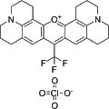
- Cat. No.
- A44994
- CAS No.
- 63561-42-2
Rhodamine 700 is a Lambdachrome laser dye characterized by its near-infrared fluorescence properties. This compound is primarily employed for mitochondrial staining in various biological research applications, enabling enhanced visualization and analysis of mitochondrial structures and functions within cells. Its unique optical characteristics make it a valuable tool in fluorescence microscopy and related studies.
-
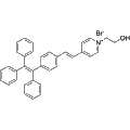
- Cat. No.
- A45047
- CAS No.
- 3032077-04-3
TPE-PY is a cationic, lipophilic fluorescent dye that targets mitochondria through the aggregation-induced emission (AIE) mechanism. Its primary mode of action involves the restriction of intramolecular rotation (RIR), resulting in enhanced fluorescence when in an aggregated state. TPE-PY is primarily utilized in biological imaging to specifically stain mitochondria in living cells, allowing for detailed observation of mitochondrial location, morphology, and abundance.
-
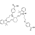
- Cat. No.
- A45285
- CAS No.
- 1895075-45-2
IR-7 is a mitochondria-targeted heptamethylindocyanine dye designed for bioimaging applications. This compound exhibits selective staining of mitochondria, making it a valuable tool for studying mitochondrial dynamics and function in various cellular contexts. Researchers can leverage IR-7 to investigate cancer biology, particularly in understanding mitochondrial roles in tumorigenesis and metabolism.
-
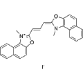
- Cat. No.
- A45342
- CAS No.
- 522592-13-8
Synonyms: D-22421
JC-9 is a green-fluorescent probe that specifically targets mitochondrial membrane potential. It enables ratiometric analysis, allowing researchers to assess changes in mitochondrial function with high sensitivity. This reagent is valuable in studies of cellular bioenergetics and mitochondrial-related diseases, facilitating a deeper understanding of mitochondrial dynamics and health.
-
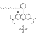
- Cat. No.
- A45352
- CAS No.
- 877933-92-1
Rhodamine B hexyl ester perchlorate is a cell-permeant fluorochrome that specifically labels mitochondria with an orange fluorescence. This compound is widely used in cellular imaging studies to assess mitochondrial function and distribution. Its fluorescence properties make it an ideal tool for visualizing mitochondrial dynamics in a variety of biological contexts.
-
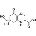
- Cat. No.
- A49021
- CAS No.
- 65318-21-0
Synonyms: Mycosporine-Gly
Mycosporine glycine is a potent singlet oxygen quencher that mitigates the harmful effects of Type-II photosensitization in biological systems. It effectively prevents mitochondrial electron transport inactivation, lipid peroxidation of microsomes, erythrocyte hemolysis, and growth inhibition in E. coli. This compound is valuable in research focused on photoprotection in marine organisms, offering insights into strategies for mitigating sunlight-induced damage.
-
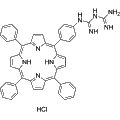
- Cat. No.
- A53952
- CAS No.
- 1014978-87-0
Biguanidinium-porphyrin is a mitochondria-targeting photosensitizer known for its ability to localize within cellular membranes, particularly within vesicles associated with lysosomes. With extended exposure, it accumulates in mitochondria and the cytosolic membrane. This compound is valuable in research applications focused on photodynamic therapy and cellular imaging, enabling investigations into mitochondrial function and dynamics.
-
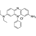
- Cat. No.
- A78599
- CAS No.
- 4569-86-2
Methylene Violet 3RAX is a phenazine dye primarily targeting mitochondria for cellular staining. This reagent can alter DNA molecular structure and induce the production of reactive singlet oxygen. It also exhibits inhibitory activity against human erythrocyte acetylcholinesterase (AChE) and human plasma butyrylcholinesterase (BChE) with inhibition constants (Ki) of 1.58 and 0.51 µM, respectively. Methylene Violet 3RAX is valuable for researching potential photosensitizers aimed at mitochondrial targeting in photodynamic therapy (PDT).
-
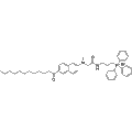
- Cat. No.
- B11256
- CAS No.
- 3091879-75-0
Mito-laurdan bromide is a fluorescent probe designed to target lipid environments within mitochondria. This compound features a cationic triphenylphosphonium moiety that selectively accumulates at the inner mitochondrial membrane due to the negative membrane potential, connected by a three-carbon linker. Mito-laurdan bromide enables the study of lipid dynamics and organization in mitochondrial membranes, making it an invaluable tool for research in bioenergetics and mitochondrial function.
-
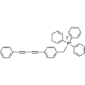
- Cat. No.
- B11312
- CAS No.
- 1644119-76-5
MitoBADY is a mitochondria-selective Raman probe designed to visualize mitochondria in live cells. At concentrations ranging from 200-400 nM, MitoBADY effectively marks mitochondria in HeLa cells for up to 10 minutes, facilitating real-time imaging. This reagent is valuable for investigating mitochondrial dynamics and studying various cellular processes related to mitochondrial function and pathology.
Selection Guide
Define the readout.
JC-1 changes between green monomer and red aggregate signals. TMRE responds through accumulation, but the cited isolated-mitochondria study also found quenching, binding and effects on respiration. Do not assume that brighter always means a higher potential. [1] [2]
Match the sample and controls.
Use the cited experimental context to plan controls and interpretation, rather than transferring a result between dyes or cell types. Green FM results depended on cell condition and removal of unbound dye before confocal imaging. [3]
Plan sample processing.
The Green FM example here concerns living cells and tissues. It does not establish a general fixed-cell workflow. Check the intended processing sequence separately; no blanket claim about aldehyde retention is made here. [3]
Representative Reagent Comparison
Selected examples for this topic; independent of the current product page or filters.
Representative reagents and selection considerations
| Reagent / SKU | Literature-supported use | Readout / interpretation | Experimental boundary |
|---|
| JC-1A15135 |
Membrane-potential-related measurements. | Green monomer and red aggregate signals; aggregation also depends on dye concentration. | The cited characterization used buffers and isolated cardiac mitochondria. Interpret both signals with appropriate controls, not as a mitochondrial count. [1] |
|---|
| TMREA24538 |
Membrane-potential assessment in isolated rat-heart mitochondria. | Potential-sensitive accumulation; fluorescence can be quenched after accumulation. | Membrane binding and respiratory effects affected interpretation. Use a TMRE-specific method; TMRM conditions are not interchangeable. [2] |
|---|
| MitoTracker Green FMA24612 |
Mitochondrial localization in living cells and tissues. | Green fluorescence; relative insensitivity to potential was conditional in the cited study. | Cell damage and unbound dye affected readings. No general fixation-retention claim or direct mitochondrial-count calibration is established here. [3] |
|---|
A15135
- Literature-supported use
- Membrane-potential-related measurements.
- Readout / interpretation
- Green monomer and red aggregate signals; aggregation also depends on dye concentration.
- Experimental boundary
- The cited characterization used buffers and isolated cardiac mitochondria. Interpret both signals with appropriate controls, not as a mitochondrial count. [1]
A24538
- Literature-supported use
- Membrane-potential assessment in isolated rat-heart mitochondria.
- Readout / interpretation
- Potential-sensitive accumulation; fluorescence can be quenched after accumulation.
- Experimental boundary
- Membrane binding and respiratory effects affected interpretation. Use a TMRE-specific method; TMRM conditions are not interchangeable. [2]
A24612
- Literature-supported use
- Mitochondrial localization in living cells and tissues.
- Readout / interpretation
- Green fluorescence; relative insensitivity to potential was conditional in the cited study.
- Experimental boundary
- Cell damage and unbound dye affected readings. No general fixation-retention claim or direct mitochondrial-count calibration is established here. [3]
These examples are not a ranking or a statement of interchangeability. Match the reagent, sample preparation and controls to the intended readout.
References
-
Reers M, Smith TW, Chen LB. J-aggregate formation of a carbocyanine as a quantitative fluorescent indicator of membrane potential.
Biochemistry. 1991. DOI: 10.1021/bi00232a015. PMID: 2021638.
-
Scaduto RC, Grotyohann LW. Measurement of mitochondrial membrane potential using fluorescent rhodamine derivatives.
Biophys J. 1999. DOI: 10.1016/s0006-3495(99)77214-0. PMID: 9876159. PMCID: PMC1302536.
-
Pendergrass W, Wolf N, Poot M. Efficacy of MitoTracker Green and CMXrosamine to measure changes in mitochondrial membrane potentials in living cells and tissues.
Cytometry A. 2004. DOI: 10.1002/cyto.a.20033. PMID: 15382028.
