Search by product name, CAS, target or catalog number. Press Down Arrow to reach product suggestions, Tab to move between links, and Escape to close suggestions.
USD – US Dollar✓Current currency
Account
Cell Viability & Live/Dead Stains
Choose a reagent by the cellular property you need to assess: membrane integrity, intracellular enzyme activity or metabolic reducing activity. These readouts answer different questions and should not be treated as interchangeable measures of survival. [1][2][3] Compare the selected reagents below, then match the assay to your sample, treatment and detection method. Use complementary measurements when a single signal cannot resolve the biological question.
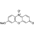
Resazurin sodium is a non-toxic, stable redox indicator that targets bacterial activity through its membrane-permeable properties. It serves as a vital tool for assessing cell viability, proliferation, and toxicity in various cell types, including human, plant, animal, bacterial, and fungal cells. Upon reduction, Resazurin sodium transforms into Resorufin, a highly fluorescent compound, enabling sensitive detection and quantification of cellular processes under fluorescent microscopy or spectrophotometry.
Calcein-AM becomes fluorescent after esterase conversion, whereas propidium iodide uptake can reveal loss of membrane exclusion. Human trabecular-meshwork experiments used both reagents with viable and damaged-tissue controls. That example supports complementary readouts, not a universal threshold for every sample. [1][2]
Could the treatment change the reporter?
Calcein retention depends on cellular conditions as well as conversion. Resazurin reports reducing activity, which can change without demonstrating cell death. Include appropriate assay controls and account for non-cellular reduction when interpreting metabolic assays. [1][3]
What conclusion is needed?
Distinguish an esterase/retention signal, a membrane-integrity observation and a metabolic comparison in your analysis. Do not rename all three as a direct count of living cells. The selected examples do not establish an apoptosis mechanism or a proliferation rate. [1][2][3]
Representative Reagent Comparison
Selected examples for this topic; independent of the current product page or filters.
Representative reagents and selection considerations
Reduction to resorufin; fluorescence or absorbance readout.
Metabolic reducing activity rather than direct death staining.
The paper supports resazurin assay principles, not SKU-specific performance. Non-cellular reduction and nonlinear response can bias interpretation. [3]
Reduction to resorufin; fluorescence or absorbance readout.
Useful distinction
Metabolic reducing activity rather than direct death staining.
Experimental boundary
The paper supports resazurin assay principles, not SKU-specific performance. Non-cellular reduction and nonlinear response can bias interpretation. [3]
Use the exact reagent form and controls appropriate to your model. The comparison is not a survival ranking or an Adooq experimental validation.